Retina & Macular Disease
The retina is the light-sensing tissue at the back of the eye, and a wide range of conditions (from diabetic retinopathy and macular degeneration to retinal tears, detachment and vessel occlusions) can threaten the vision it makes possible.
What is the retina? Where is it and what does it do?
The retina, the net-like layer, is a vital sheet of tissue at the back of the eye, rich in blood vessels and nerve cells, that senses light and begins the process of vision. About as thin as a fine business card, the retina is made up of light-sensitive cells (photoreceptors) and nerve cells. The light that enters our eye is processed by these cells in the retina and carried to the brain as visual signals through the optic nerve. In short, the retina works as the eye's "vision center."
The retina has two main jobs. First, sensing light: the retina receives the light focused by the eye's lens and cornea and converts it into nerve signals: the very first step of seeing. Second, carrying visual signals to the brain: the retina transmits these signals through the optic nerve, and the brain processes them to form the image, so that vision takes place.
Two kinds of cells do this work. Rods provide the retina's sensitivity to light; they make vision possible especially in low-light conditions and are important for peripheral vision. Cones provide color vision; they work harder in bright light and are responsible for sharp, central vision.
The retina is the most important part of the eye for carrying the perception of light and color to the brain. Any problem or disease in the retina can cause serious vision loss, so retinal health is critically important for preserving the ability to see.
Several factors affect retinal health. With aging, retinal function can deteriorate; conditions such as age-related macular degeneration in particular can harm the retina, because the oxidation that comes with aging triggers damage to nerve cells, the cells most sensitive to oxygen deprivation, and leads to various retinal diseases. Diabetes can cause vision loss by damaging the retina's blood vessels; it is a disease that affects the small vessels and also causes nerve damage (neurodegeneration), so the retina, rich in both nerve and vascular cells, is the tissue most affected by it. Eye injuries: physical trauma can lead to retinal damage and serious vision loss. Genetics: many retinal diseases have a genetic basis, and people with familial retinal disease should not neglect their eye check-ups. Systemic diseases: various conditions in the body affect the retina: vascular diseases such as hypertension and atherosclerosis, rheumatic diseases, and the use of certain medications can all damage the retina in particular.
As we always tell our patients, a healthy retina is found in a healthy body. Anything that harms the body (smoking, poor nutrition, stress) or many diseases (the common ones being diabetes, hypertension and atherosclerosis) can affect retinal health and therefore vision.
What is the macula (the "yellow spot")?
The macula is the part of the retina at the back of the eye that provides central vision. It is present in every person. The macula is the region of the retina that makes clear, detailed vision possible and the place where light is focused. It plays a critical role in activities that require fine vision, such as recognizing faces, reading and writing. The fovea lies right at the center of the macula, and the sharpest vision happens there. The macula is called the "yellow spot" because it is an area dense in pigment, and it is vital to the healthy working of the retina. It is very vulnerable to oxygen deprivation, so if the oxidant–antioxidant balance in the body and retina is disturbed, the macula is affected and various diseases appear.
The main macular conditions are: damage and breakdown of the macula (age-related macular degeneration); swelling in the macula (macular edema due to diabetes or to retinal vein occlusion); a hole in the macula (macular hole); a membrane over the macula (epiretinal membrane); and inherited macular diseases (Stargardt disease, cone dystrophy, retinitis pigmentosa).
Symptoms of retinal disease and early diagnosis
The retina is the important layer at the back of the eye that senses light and begins the process of vision. Any disease or damage to the retina can cause vision loss. Recognizing the early signs of these diseases and intervening in time is critically important for preventing permanent vision loss.
The common symptoms of retinal disease include the following. Blurred vision is the most common sign, seen especially in diabetic retinopathy, macular degeneration or retinal detachment. Flashes of light, sudden and frequent, can herald a retinal tear or detachment and require urgent attention. Floaters (floating dots, threadlike shapes or shadows in the field of vision) are one of the early signs of retinal disease and can arise when the vitreous fluid presses on the retina; with age such symptoms can sometimes appear normally too, but an eye examination is needed to tell the difference. Distorted central vision: because of macular (central retina) problems, central vision may blur and straight lines may look bent or wavy, which can be a sign of age-related macular degeneration. Seeing objects as crooked, broken, or larger or smaller than they are can be caused by age-related macular degeneration or by other diseases affecting the macula. Loss in the field of vision or blind spots: retinal detachment, diabetic retinopathy or vessel occlusions can produce dark patches or blind spots. Colors looking faded: retinal diseases can make colors appear less vivid and washed out, a symptom that is common especially in macular degeneration patients.
Why is early diagnosis important? If retinal diseases are diagnosed and treated in their early stages, vision loss can be prevented or at least slowed. However, because many retinal diseases show no obvious symptoms at first, regular eye examinations are very important.
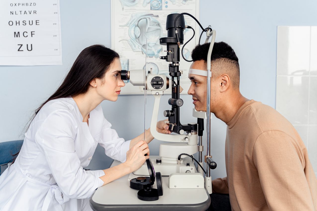
Early diagnosis is possible through several methods. Fundus examination (fundoscopy) lets the retina be examined directly so that possible tears, vessel occlusions or other damage can be detected; this is performed by eye doctors. Optical coherence tomography (OCT) is used to image the retinal tissues in detail and plays a major role in diagnosing macular degeneration, diabetic retinopathy and other retinal diseases. Fluorescein angiography is used to identify problems in the retina's blood vessels and shows whether there is leakage or blockage. The Amsler grid test is a simple test used to detect whether central vision is distorted; with it, patients check whether straight lines appear wavy or broken.
Some recommendations for early diagnosis: have regular eye check-ups, especially as the risk of retinal disease is known to rise in people aged 40 and over: an eye examination at least once a year is important for early detection. Pay attention to family history, since some retinal diseases (such as retinitis pigmentosa) can be inherited, and people with eye disease in the family should be more careful. Control diabetes and hypertension, because diseases such as diabetic retinopathy are linked to diabetes and high blood pressure; keeping blood sugar and blood pressure under control is an important step in protecting the retina, and people with these conditions need regular eye check-ups.
Diagnostic methods for retinal disease
Early diagnosis of retinal disease is very important for preventing or slowing vision loss. Here are the methods we use most often.
Optical coherence tomography (OCT). How does it work? It is an imaging test, the one most used by eye doctors who focus on the retina, that creates detailed images of the retinal tissue. OCT is used to examine the retinal layers and to diagnose problems such as fluid accumulation and retinal tears. It is common in diagnosing diabetic retinopathy, macular degeneration and glaucoma. Its advantage is that, by providing cross-sectional images of the retinal tissue, it can detect even the smallest changes in the retina, almost as if a pathological or histological sample had been taken.
Fluorescein angiography. How does it work? This test uses a special dye (fluorescein) and a camera to examine the retina's blood vessels. Conditions such as vessel occlusions, bleeding and leakage are detected by this method. Its advantage is that it clearly shows the structure of the retinal vessels and the blood circulation; it is used in diseases such as diabetic retinopathy and macular degeneration.
Fundoscopy (fundus examination). How does it work? The eye doctor uses a special lens and light to examine the retina at the back of the eye. The fundus examination is the most basic method for detecting retinal disease. Its advantage is that it allows direct observation of the retinal vessels, the optic nerve and the macula.
Amsler grid test. How does it work? It is a simple test used to detect distortions in the central field of vision. It is common in diagnosing diseases that cause central vision loss, such as macular degeneration. Its advantage is that it is a test the patient can even apply at home, and it gives an early warning.
Electrophysiological tests (ERG and VEP). How do they work? These tests measure the electrical activity of the retinal cells and the optic nerve. They are used in diagnosing rare retinal diseases such as retinitis pigmentosa. Their advantage is that they detect functional disorders in the retina and the visual pathways.
OCTA (optical coherence tomography angiography). How does it work? OCTA is a test that creates a detailed image of the retinal and choroidal vessels. Unlike traditional fluorescein angiography, no dye is used; it maps the retinal vessels by measuring the reflection of light from the moving blood cells in the retina. Its advantages and uses: it is used in diagnosing diseases that especially affect the macula, such as diabetic retinopathy, age-related macular degeneration and retinal vessel occlusions. It is an extremely sensitive and innovative method for diagnosing retinal disease.
Retinal surgery: types, risks and the recovery process
Retinal operations are critical surgical methods used in treating retinal disease. Because retinal damage or disease often requires urgent intervention, these operations are vital for preventing serious vision loss.
Vitrectomy (pars plana vitrectomy, vitreoretinal surgery). How is it done? The vitreous (the gel inside the eye) is removed and retinal damage is repaired. It is used to treat retinal tears, detachment or bleeding. Uses: retinal detachment, diabetic retinopathy, macular hole. Risks: infection, bleeding, development of cataract.
Pneumatic retinopexy. How is it done? A gas bubble is injected into the eye and the retina is fixed back into place. It is generally used for retinal tears and small detachments. Uses: small retinal tears or localized detachments. Risks: the gas bubble not settling fully, vision disturbance.
Scleral buckling. How is it done? A silicone band is placed on the outside of the eye and the retinal damage is repaired. It is a common surgical method for treating retinal detachment. Uses: advanced retinal detachments. Risks: distortion of the eye's shape, infection.
Laser treatment (photocoagulation). How is it done? Laser is applied around the retinal tears so that the tears are "sealed" and retinal detachment is prevented. Uses: retinal tears, abnormal growth of blood vessels. Risks: it is minimally invasive, but it can rarely cause mild blurring of vision.
The risks of these operations include the following. Infection: as with every surgical procedure, there is a risk of infection. Bleeding: bleeding may occur in the retinal area during or after surgery. Cataract development: this is a common complication after vitrectomy. Vision loss: one of the risks of surgery is vision loss, though it is rare.
The recovery process: the recovery time after surgery can take from a few weeks to a few months depending on the operation performed. Heavy physical activity should be avoided during the first few weeks. In operations that use a gas bubble, the patient's head position is very important; following the doctor's instructions, the patient may need to lie in a particular position. Vision may not improve immediately after surgery; full recovery can take several weeks.
Post-operative care: eye drops: antibiotic and steroid drops are prescribed after surgery to reduce the risk of infection and speed recovery. Regular check-ups: regular eye examinations after surgery are important for assessing recovery and preventing complications.
What are intraocular injections (needle treatment)?
Intraocular injections are an effective treatment method used in retinal and macular diseases. This treatment is done by injecting medication directly into the eye, providing a fast and direct effect on the retinal area. Medicines given by mouth or through a vein have limited effect on the retina because of the blood–retina barrier, and eye drops also cannot fully pass through the cornea and sclera surrounding the eye to reach the retina at the back. The most commonly used medicines are anti-VEGF (vascular endothelial growth factor inhibitors) and steroids. Unfortunately, the healing effect of both treatments is temporary: anti-VEGF agents generally last 1–3 months and steroids (dexamethasone implant) 3–4 months. Sometimes this period can be longer, or, on the contrary, even shorter in severe disease.
Where is it used? Age-related macular degeneration (AMD), diabetic retinopathy, retinal vessel occlusions, and other vascular diseases of the retina.
The treatment process: the procedure is generally quick and is done with drop anesthesia. After the medicine is injected into the eye, it acts on the retina, stopping vascular leakage and slowing vision loss. After treatment, patients may feel mild discomfort but usually recover quickly; improvements in visual function become noticeable after a few injections.
Risks: infection, a rise in intraocular pressure, or bleeding in the eye can occur, though rarely. These risks are kept under control with regular follow-up examinations.
Follow-up and afterward: rarely, a single injection is enough in some patients, but in the great majority of patients regular injections are needed. The first three injections are given monthly. After that there are various treatment regimens, and treatment continues with the injection need determined for each patient individually. Regular check-ups are very important, because in high-risk lesions, if an injection is delayed, the vascular lesions can bleed and cause permanent vision loss.
Recommendations for protecting retinal health
Protecting retinal health is critically important for overall eye health. Some important steps can be taken to prevent or delay retinal disease.
Balanced nutrition. Antioxidants such as lutein and zeaxanthin protect the retina against oxidative stress; green leafy vegetables such as spinach, kale and broccoli support retinal health. Omega-3 fatty acids (foods rich in omega-3 such as fish (salmon, tuna)) protect the retinal cells and reduce the risk of age-related macular degeneration. Vitamins C and E, which have antioxidant properties, protect the retina from age-related damage; citrus fruits, nuts and seeds are rich in them.
Regular exercise. Regular physical activity increases blood circulation and provides oxygen to the retinal cells. It helps control diabetes and hypertension, which in turn reduces the risk of diabetic retinopathy.
UV protection. Wearing sunglasses: long exposure to the sun can cause retinal damage, and by using UV-protective sunglasses you can minimize the harmful effects of the sun. When the eyes are not protected from UV rays, the risk of age-related macular degeneration and cataract increases.
Limiting smoking and alcohol. Smoking is one of the factors that threaten retinal health; smoking causes oxidative stress and damages the retinal vessels, and quitting contributes positively to retinal health. Alcohol: excessive alcohol consumption can damage the retinal cells, so limiting alcohol intake is important for protecting eye health.
Regular eye examinations. Retinal diseases may show no symptoms in their early stages, so going to the eye doctor at least once a year is critically important for protecting retinal health. People with risk factors such as diabetes and hypertension in particular need regular examinations.
Control of diabetes, hyperlipidemia and hypertension. High blood pressure and high blood sugar levels can damage the retinal vessels. Keeping these health problems under control is important for preventing diseases such as diabetic retinopathy and hypertensive retinopathy.
Supplements for eye health. Eye-health supplements containing lutein, zeaxanthin and vitamins C and E may help protect the retina; such supplements may in particular help prevent age-related retinal damage.
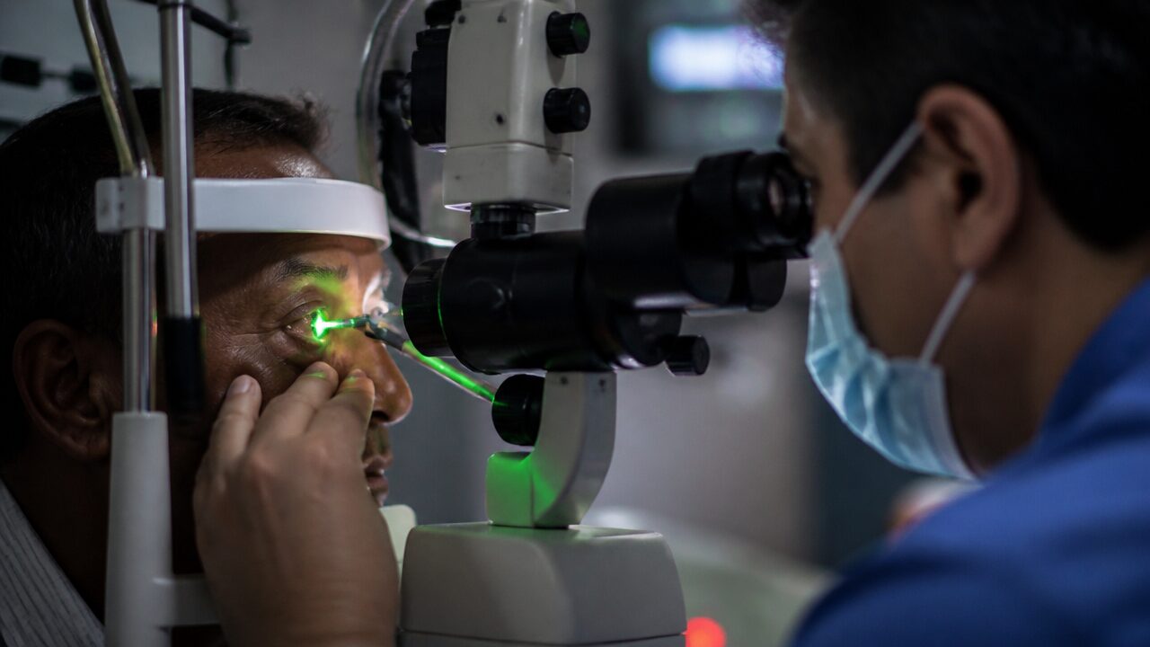
Laser treatments of the retina and argon laser photocoagulation
Argon laser photocoagulation is a type of laser used in treating retinal disease. By applying a laser beam to specific areas of the retina, abnormal vessels are closed, leakage is stopped, or the retinal tissue is made to reattach.
How does laser work on the retina? The argon laser gives heat to the small blood vessels in the retina so that these vessels close. By applying laser around retinal tears, it "welds" and seals the edges of the tears and prevents retinal detachment.
Where is retinal laser used? In diabetic retinopathy, it stops abnormal vessel growth. In retinal tears and detachment, it seals around the tears. In retinal occlusions, it controls blood leakage and edema.
Advantages: it is a minimally invasive method, can be done on an outpatient basis, and can slow or stop vision loss. Side effects of retinal laser: mild pain and discomfort, temporary blurred vision, rarely a reduction in the field of vision, and disturbances in night vision.
Panretinal argon laser is applied to the entire retina over a few sessions, sparing the macula and its surroundings. Focal laser, on the other hand, is applied to the leaking microaneurysms (small vascular blisters) around the macula. This treatment can generally be applied over several sessions and is an effective way to halt the progression of retinal disease.
What is a retinal tear? Symptoms and treatment
A retinal tear is damage to the retinal layer at the back of the eye, which is critical for the function of vision. The retina is a tissue tightly attached to the inner wall of the eye, and it senses light and carries visual information to the brain. When the retina tears, fluid can seep beneath it and cause the retinal layer to separate from its place (retinal detachment). This situation requires urgent attention.
Causes of a retinal tear: a tear generally occurs when the vitreous (the gel-like substance inside the eye) presses on or pulls at the retina. With age, the vitreous fluid loses volume inside the eye and, shrinking, can stick to the retina. Other factors that can lead to a tear include aging (with age the vitreous fluid can separate from the retina), eye injuries (traumatic eye injuries can cause a retinal tear), high myopia (the risk of a retinal tear is increased in people with high myopia) and previous eye surgery (the risk can rise especially after cataract surgery).
The seriousness of a retinal tear: if untreated, a retinal tear can lead to retinal detachment (the retina separating completely from its place). This can cause serious and permanent vision loss, which is why retinal tears require urgent treatment.
Symptoms of a retinal tear: sudden flashes of light (a flash or lightning sensation in the eye), floaters in the eye, and dark shadows or a curtain over the field of vision.
Treatment of a retinal tear: laser treatment (laser photocoagulation): laser is applied around the retinal tear so that the borders of the tear are "welded" and the seeping of fluid beneath the retina is prevented. Cryotherapy: the retinal tear is frozen so that the tissues around it become adherent, which prevents fluid from seeping beneath the retina. Surgical intervention: in advanced retinal detachment cases, surgical procedures such as vitrectomy are applied; vitrectomy is the removal of the vitreous gel from the eye and the repair of the retinal tear.
The importance of early intervention: it is critical to see an eye doctor as soon as the symptoms of a retinal tear are noticed. Early diagnosis and treatment can prevent the development of retinal detachment and permanent vision loss. People who notice sudden flashes of light or floaters in particular should have an urgent eye examination.
What is retinal detachment?
The retinal layers formed by the retina's nerve cells are attached to a support layer called the epithelium, much like wallpaper stuck to a wall. Retinal detachment is a serious eye condition in which the retina separates from these support tissues beneath it, just as wallpaper peeling away from a wall. The retina is a thin layer of tissue inside the eye that senses light and forms the visual signals carried to the brain. Its separation from the tissues beneath it causes a loss of nourishment and oxygen, leading the retina to lose its function. From the moment the retina begins to separate, loss of nerve cells begins. If untreated, it can cause permanent vision loss.
Causes of retinal detachment: a retinal tear: when the retina tears, the vitreous fluid inside the eye can seep beneath it and lift it from its place. Vitreous separation: the vitreous (the gel in the eye) can pull away from the retina with age and lead to a tear. Trauma: eye injuries can cause a retinal tear or detachment. Other factors: high myopia, diabetic retinopathy and previous eye surgery (most often cataract surgery) can increase the risk of retinal detachment.
Symptoms of retinal detachment: sudden flashes of light, floaters in the field of vision, dark shadows or a curtain coming down over the field of vision, and distortion or loss of vision.
Treatment methods for retinal detachment: laser treatment or cryotherapy: in the early stages of retinal detachment, laser is applied around the retina to fix the area around the tear, or the tear is frozen and fixed with cryotherapy. Surgical intervention: in advanced cases, surgical procedures such as vitrectomy (vitreoretinal surgery, pars plana vitrectomy) or scleral buckling are applied; with these methods the retina is fixed back into place.
The importance of urgent intervention in retinal detachment: retinal detachment is a condition requiring urgent attention. Early diagnosis and treatment can prevent vision loss. When sudden changes are noticed in the eye, an eye doctor should be seen without losing time, and treatment should be started as soon as possible.
What is diabetic retinopathy? Retinal disease linked to diabetes
Diabetic retinopathy is an eye disease seen in people with diabetes, and it arises as a result of damage to the retina's blood vessels. The retina is the light-sensing tissue layer at the back of the eye. In people with diabetes, high blood sugar can damage the retinal vessels and lead to vision loss. Diabetic retinopathy is one of the most common causes of blindness in adults worldwide.
Types of diabetic retinopathy. Non-proliferative diabetic retinopathy (NPDR) is the first stage and generally shows no obvious symptom; microaneurysms and small bleeds form in the retinal vessels, and vision loss is usually minimal. Proliferative diabetic retinopathy (PDR) is the advanced stage of the disease; abnormal new vessels form on the retina, and these vessels can bleed and cause vision loss, while the risk of retinal detachment also rises.
Symptoms of diabetic retinopathy: blurred vision, dark or empty areas in the field of vision, sudden vision loss, and floaters.
Risk factors for diabetic retinopathy: the duration of diabetes (the risk rises in people who have had diabetes longer), poor blood sugar control, high blood pressure and cholesterol levels, and pregnancy (the risk rises in women with diabetes).
Treatment methods for diabetic retinopathy: laser treatment: laser is used to prevent leakage from the retinal vessels. Anti-VEGF injections: medication injections that stop the growth of abnormal blood vessels. Vitrectomy: if there is bleeding in the eye or retinal detachment, surgery is applied to remove the vitreous fluid.
Prevention of diabetic retinopathy: keeping blood sugar, blood pressure and cholesterol levels under control can slow the development of diabetic retinopathy. Having regular eye examinations is critical for early diagnosis and treatment; after a diagnosis of diabetes in particular, it is important to have an eye examination at least once a year.
The importance of early diagnosis in diabetic retinopathy: diabetic retinopathy often gives no symptoms in its early stages, so regular eye examinations are important. Early diagnosis and intervention can prevent permanent vision loss.
Eye diseases linked to diabetes: how diabetes affects the eye
Eye diseases linked to diabetes develop because of the damage that long-standing diabetes or high blood sugar levels cause to the vessels in the eye. Leaving diabetes uncontrolled can have serious effects on critical structures such as the retina, the lens and the optic nerve. Here are the eye diseases most commonly seen in relation to diabetes.
Diabetic retinopathy. What is it? It is a disease that develops as a result of diabetes damaging the retina's vessels; the retina is the light-sensing tissue at the back of the eye, and this disease can cause vision loss. Symptoms: floaters, blurred vision, dark areas or vision loss. Treatment: laser photocoagulation, anti-VEGF injections, vitrectomy.
Diabetic macular edema. What is it? The macula is the retinal region responsible for central vision. Diabetic macular edema occurs when fluid accumulates in the macula as a result of leakage from the retinal vessels, and it can lead to a serious reduction in visual acuity. Symptoms: blurred vision, central vision loss, straight lines appearing wavy. Treatment: anti-VEGF injections, laser treatment.
Cataract. What is it? Cataract arises from clouding of the eye's lens, and people with diabetes have a higher risk of developing it; the lens loses its transparency, preventing light from reaching the retina properly. Symptoms: blurred or hazy vision, sensitivity to light, inability to see clearly in low light. Treatment: in cataract surgery, the clouded lens is removed and an artificial intraocular lens is placed.
Glaucoma. What is it? Glaucoma arises from damage to the optic nerve as a result of increased intraocular pressure, and diabetes is a risk factor for its development; when the optic nerve is damaged, peripheral vision loss occurs. Symptoms: narrowing of the field of vision, headache, eye pain, blurred vision. Treatment: medications, laser treatment, surgical intervention.
Retinal detachment. What is it? Retinal detachment occurs when the retinal layer separates from the back wall of the eye; in proliferative diabetic retinopathy, retinal detachment can occur when the new vessels that form tear and bleed. Symptoms: sudden flashes of light, floaters, dark shadows or a curtain over the field of vision. Treatment: retinal detachment generally requires urgent surgical intervention.
Ischemic optic neuropathy. What is it? When the blood circulation in the eye is disturbed because of diabetes and the optic nerve does not receive enough blood, ischemic optic neuropathy can develop. This can cause sudden and permanent vision loss. Symptoms: suddenly developing vision loss, usually in one eye. Treatment: there is no definitive treatment for this condition, but keeping diabetes and blood pressure under control reduces the risks.
Eye-muscle palsies, paralytic strabismus. What is it? It is the weakening or loss of function of the eye muscles because the muscles, and the nerves that control them, fed by the small vessels blocked by diabetes, cannot be nourished. Symptoms: double vision, deviation of the eyes, restriction of eye movement in certain directions. Treatment: keeping diabetes and blood pressure under control reduces the risks; if it persists after 6 months of follow-up, surgery may be applied.
Prevention of diabetic retinopathy
Prevention of diabetic retinopathy is the most important measure for people with diabetes to protect their eye health. Here are some important steps that can be taken to prevent or slow the development of the disease.
Blood sugar control. Regular blood sugar monitoring: keeping blood sugar continuously under control plays a major role in preventing the high sugar levels that can damage the retina; keeping the HbA1c level below 7% is critical for protecting eye health. Diet: a low-sugar, fiber-rich diet should be preferred, and diet plans tailored to people with diabetes can improve eye health.
Blood pressure and cholesterol control. High blood pressure and cholesterol can damage the retinal vessels, so it is important to keep blood pressure below 140/90 mmHg. In addition, diet and medication can be used to lower bad cholesterol (LDL) levels.
Regular eye examinations. People with diabetes should have a comprehensive eye examination at least once a year. Because the signs of diabetic retinopathy are not noticed in the early stage, regular examinations are critically important for halting the progression of the disease.
Quitting smoking. Smoking can damage the retina by narrowing the blood vessels. For people with diabetes, quitting smoking significantly reduces the risk of retinopathy.
Physical activity. Regular exercise helps maintain blood sugar control and reduces the complications of diabetes; physical activity is beneficial for eye health as well.
Balanced use of medication. Using the medications recommended by the doctor regularly both keeps diabetes under control and protects eye health. Blood-sugar-regulating medicines, blood-pressure medicines and cholesterol-lowering medicines can reduce the risk of retinopathy. A regular sleep pattern and stress control are also helpful.
What is macular disease (macular degeneration)?
The macula, the yellow spot, is the part of the retina present in everyone that provides sharp, central vision. When we speak of macular diseases, we are speaking of dozens of conditions, but in particular "age-related macular degeneration," seen in individuals over 55 and leading to central vision loss as a result of damage to the macula, is the common and most widely known retinal disease among the public. The macula is the region of the retina that gives us clear, detailed vision. Progression of the disease distorts central vision and makes daily activities difficult. It is also known as age-related macular degeneration (AMD).
Types of age-related macular degeneration. Dry-type macular degeneration is the most common form of the disease (about 80% of patients) and progresses slowly; it occurs as the macula thins through the gradual degeneration of the retinal cells, and small yellow deposits called drusen that accumulate beneath the retina cause vision loss. In the dry type, patients generally experience gradual central vision loss, and in more advanced stages serious vision loss can occur. Wet-type macular degeneration is less common but leads to faster and more serious vision loss; abnormal blood vessels grow beneath the retina, and these vessels leak and damage the macula, which can cause rapid vision loss and permanent harm, with bleeding and serum leakage from the diseased vessels leading to reduced vision.
Symptoms of age-related macular degeneration: central vision loss: the disease especially affects central vision, and you may feel blurring, a dark patch or gaps in the center of your vision. Straight lines appearing bent: because of damage to the macula, straight lines may look wavy or curved. Distorted color perception: colors may appear paler and less distinct. Gradual vision loss: while the dry type generally progresses slowly, the wet type can develop suddenly.
Causes and risk factors of macular disease: age: the risk rises in people aged 50 and over. Genetic factors: the risk is higher in people with a family history of macular disease. Smoking: the risk of the disease doubles in smokers. Obesity and hypertension: these conditions can speed the development of macular disease. Sun exposure: long exposure to UV rays can lead to retinal damage.
Treatment methods for age-related macular degeneration: anti-VEGF injections: in the wet type, this is treatment by intraocular injection to stop the growth of abnormal blood vessels; anti-VEGF (vascular endothelial growth factor inhibitor) drugs slow vision loss by preventing the leakage and growth of blood vessels. Vitamin and mineral supplements: for the dry type, studies show that certain vitamins (vitamins C and E, beta-carotene, zinc and copper) slow progression, and vitamin-and-mineral supplements called AREDS can slow the progression of the disease. Photodynamic therapy: used in the wet type, this method uses a combination of a light-sensitive drug and laser to close the vessels.
Preventive measures: balanced nutrition: consuming foods rich in antioxidants such as lutein and zeaxanthin (green leafy vegetables such as spinach and kale) can contribute to eye health; increasing omega-3 intake, a diet rich in antioxidant foods such as walnuts, flaxseed and broccoli, olive oil instead of solid fats, and white meat instead of red meat are recommended. Quitting smoking: smoking accelerates macular degeneration, and quitting greatly reduces the risks. Wearing sunglasses: using UV-protective sunglasses outdoors to minimize exposure to UV rays is important for retinal health.
Progression and prognosis: macular disease, especially age-related macular degeneration, is a progressive condition. While the dry type progresses slowly, the wet type can progress much faster and cause serious vision loss. Early diagnosis is very important for the success of treatment.
Retinal vessel occlusions: retinal vein and artery occlusions
Retinal vessel occlusions are eye diseases that develop when the blood vessels in the retina become blocked for various reasons. This blocks blood circulation in the eye, and when enough oxygen and nutrients do not reach the retinal tissue, it can lead to serious vision loss. Nerve cells are very vulnerable to oxygen deprivation, and if a vessel occlusion does not open within a short time, retinal damage occurs and causes vision disturbances. There are two main types.
Central retinal vein occlusion (CRVO). What is it? It occurs when the central retinal vein, the main vessel leaving the retina, becomes blocked. This blockage causes the retinal vessels to leak blood and the retinal tissue to swell. Symptoms: sudden and painless vision loss, blurred vision, bleeding in the eye. Risk factors: high blood pressure, diabetes, glaucoma, cholesterol, blood disorders, and advanced age. Treatment: it is treated with anti-VEGF injections, laser treatment and corticosteroid injections; if untreated it can cause permanent vision loss.
Central retinal artery occlusion (CRAO). What is it? It occurs when the main artery that supplies oxygen to the retinal cells becomes blocked. This stops blood flow to the retina and causes sudden vision loss. Symptoms: sudden and usually painless vision loss, sometimes dark areas in the field of vision. Risk factors: cardiovascular diseases, hardening of the arteries, hypertension, diabetes, high cholesterol. Treatment: it requires urgent intervention, but in many cases permanent vision loss can occur; treatments that lower intraocular pressure may be applied, and in recent times hyperbaric oxygen treatments are being used.
Branch retinal vein occlusion (BRVO). What is it? It occurs when one of the smaller vessels of the retina becomes blocked. This blockage prevents blood from leaving the retina and causes the retinal tissue to swell. Symptoms: dark areas or blurred vision in the sight, floaters in the eye. Risk factors: high blood pressure, diabetes, glaucoma, smoking. Treatment: laser treatment, anti-VEGF injections and corticosteroids.
Branch retinal artery occlusion (BRAO). What is it? It occurs when one of the small arteries of the retina becomes blocked. This stops blood flow to the area of the retina fed by the blocked vessel. Symptoms: sudden and painless vision loss, usually loss in a part of the field of vision. Risk factors: atherosclerosis (hardening of the arteries), diabetes, high blood pressure. Treatment: treatment is generally limited, and in most cases permanent vision loss can occur; treatments that lower intraocular pressure are applied to try to restore blood flow in the eye.
General risk factors for retinal vessel occlusions: high blood pressure (hypertension), diabetes, high cholesterol, smoking, glaucoma, atherosclerosis (hardening of the arteries), and certain blood disorders that increase blood clotting.
Treatment and prevention: anti-VEGF injections stop abnormal vessel development in the retina and prevent fluid accumulation. Laser treatment is used on the blocked vessels to prevent blood leakage. Corticosteroid injections reduce inflammation and swelling in the retina. Lifestyle changes: bringing conditions such as high blood pressure, diabetes and cholesterol under control is important for preventing retinal vessel occlusions.
In macular disease, will I have injections in my eye continually?
Retinal vessel occlusions can often cause sudden and serious vision loss. Early diagnosis and treatment are very important for preventing permanent harm. In treating macular disease (age-related macular degeneration: AMD), intraocular injections are frequently used, especially in treating the wet type. These injections contain anti-VEGF drugs that prevent the growth of abnormal blood vessels and slow vision loss by reducing fluid accumulation beneath the retina.
Will injections be given in the eye continually? The injections used in treatment are generally applied at regular intervals, because the effect of the medicines lasts only a limited time. Treatment can vary depending on the speed of the disease's progression, the response to treatment, and the eye doctor's assessment. At the start an injection may be given once a month, and during this time the effectiveness of the treatment is observed. If the treatment is successful, the frequency of injections can be reduced according to the doctor's decision. In some patients we can extend this interval up to 4 months. But you should not forget this: in some patients we have given dozens, even perhaps 100, injections in the eye for macular disease, while in others a few injections are enough. This depends on the type of the disease, its course and many variables.
The anti-VEGF treatment process: in the first period, generally one injection is given per month. Depending on the response, if the patient's vision improves or stabilizes, the intervals between injections can be lengthened (for example, every two or three months). Long-term follow-up: in some patients treatment can become ongoing, but the treatment process can be different for each patient; because the effect of the medicines is short-lived, regular injections may be needed to keep the disease from progressing. Because the treatment process is shaped by the speed of the disease's progression and each individual's response, it is very important to follow your doctor's recommendations and to attend regular eye check-ups. Please do not neglect your follow-up and treatment.
Membrane over the macula (epiretinal membrane)
An epiretinal membrane (ERM) is the formation of a thin membrane over the macula. This membrane generally develops with age and forms as a thin layer on the surface of the macula. The macula is the region of the retina known as the yellow spot and is responsible for central vision. An epiretinal membrane can affect vision in this region and lead to blurred vision, fluctuating vision or distorted vision.
How it forms and its relation to macular disease: an epiretinal membrane usually develops after a small injury or trauma in the retina. As the body tries to repair this damage, a new membrane can form on the retinal surface. This membrane creates tension over the macula and causes vision disturbance. While this tension-creating membrane is thin at first, it may not give any symptom; but over time, by pulling on the macula, it distorts its shape, or by thickening it reduces the light reaching the macula and therefore vision.
Symptoms: images appearing distorted, wavy or blurred, distortion of central vision, and reduced visual acuity. An epiretinal membrane generally progresses slowly and gives very few symptoms in the early stages, but as it advances the tension over the macula increases and vision problems become more obvious.
Treatment: observation: in mild cases, if the vision disturbance is not very serious, it can be monitored with regular follow-up. Surgical intervention (vitrectomy): if the epiretinal membrane leads to significant vision loss, the membrane can be removed with a surgical procedure called vitrectomy; in this operation, the membrane over the macula is peeled away to try to improve visual function. This is an operation that requires experience and can be performed by a vitreoretinal surgeon. An epiretinal membrane can generally appear in older age or as a result of other diseases in the eye (diabetic retinopathy, retinal tears, etc.). Treatment varies depending on the thickness of the membrane and the degree of the patient's vision loss.
Hole in the macula (macular hole)
A macular hole is a small tear or hole that forms in the macular region (the yellow spot) of the retina. The macula is the retinal region responsible for central vision and is at the center of our ability to see detail. A macular hole can seriously affect this central vision and, progressing over time, can lead to vision loss.
Why does a macular hole form? Vitreous pulling: with age, the vitreous fluid, the gel-like substance inside the eye, begins to shrink and separate from the retina; during this separation the vitreous can pull on the macula and create a tear or hole. Eye injuries: trauma or injury to the eye can cause a hole to form in the macular region. Retinal diseases: other retinal diseases or retinal detachments can also lead to a macular hole. Idiopathic: sometimes a macular hole can form with no obvious cause, especially in older age.
Symptoms of a macular hole: central vision loss: a blurred or dark spot forms in the center of vision, which is usually the first sign noticed. Straight lines appearing bent: because of structural changes in the macular region, straight lines look wavy or curved. Difficulty seeing detail: you may have trouble reading small print, recognizing faces or seeing fine details.
Stages of a macular hole: macular holes develop in three stages. In Stage 1 there is only mild central vision loss and the hole has not fully formed. In Stage 2 the hole forms completely and central vision is noticeably distorted. In Stage 3 the hole enlarges to affect a large part of the macula and serious central vision loss occurs.
Treatment of a macular hole: vitrectomy: the most common surgical method in treating a macular hole is the vitrectomy operation; in this procedure, the vitreous gel inside the eye is removed and a gas bubble is injected in its place, and the gas bubble applies pressure to the retina to close the macular hole. Gas bubble: after surgery the patient may need to keep the head in a particular position; the gas bubble is gradually absorbed in the eye and replaced by eye fluid. Recovery process: visual improvement after surgery can take several months; in some cases full recovery may not be achieved, but vision loss is generally reduced.
Risks related to a macular hole: age: a macular hole is generally seen more often in individuals over 60. Eye injuries: the risk rises in people who have had an eye injury in the past. A macular hole in the other eye: if a macular hole has formed in one eye, there is a risk of development in the other eye. A macular hole is a serious condition, but with early diagnosis and treatment central vision loss can be halted or improved.
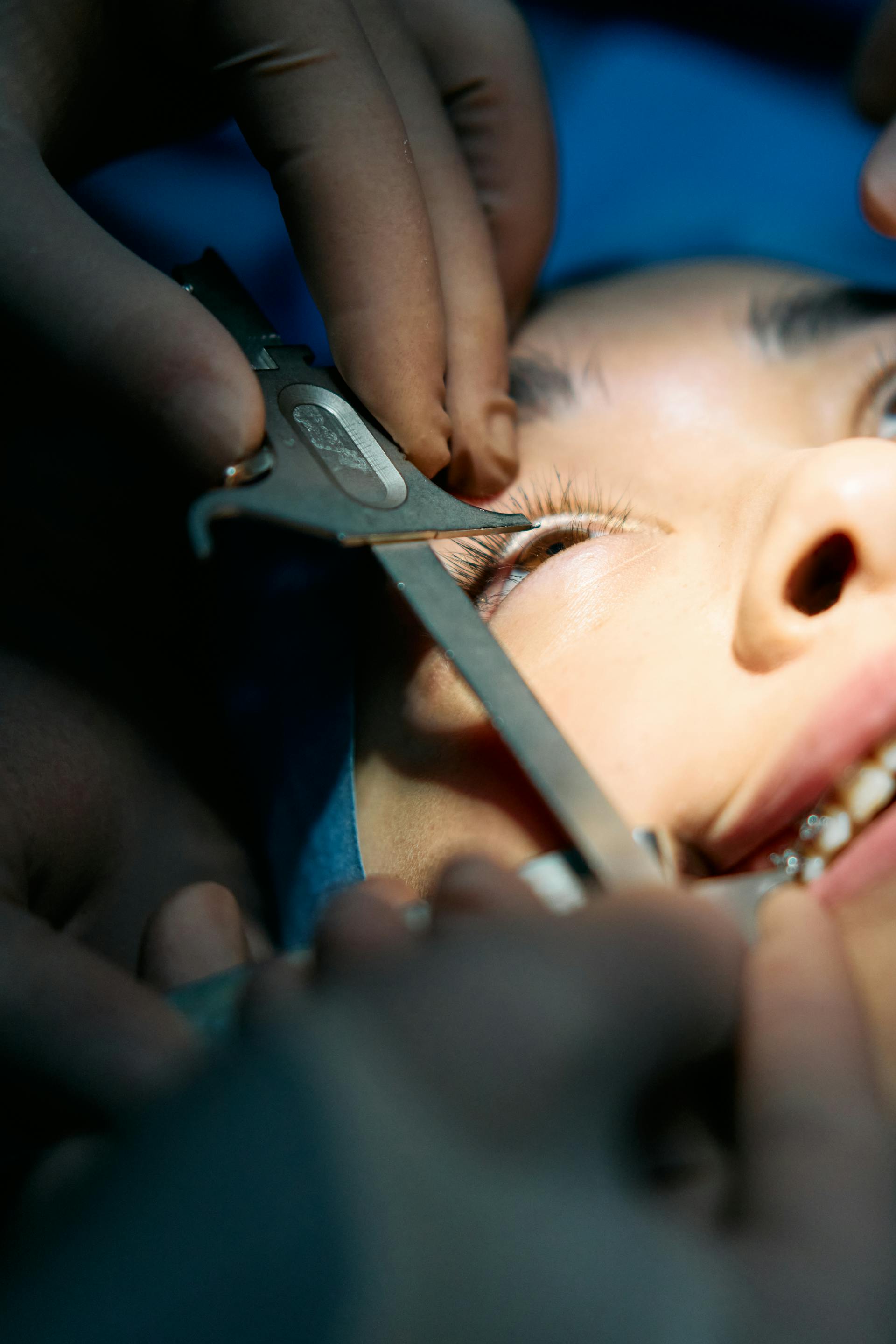
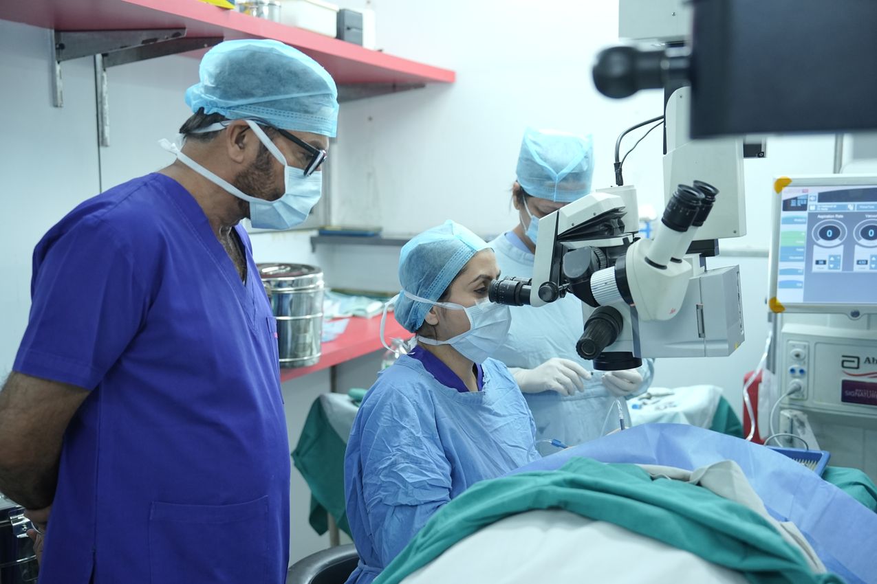
Vitrectomy, pars plana vitrectomy, vitreoretinal surgery
Vitrectomy is a surgical procedure in which the vitreous gel, the gel-like substance inside the eye, is removed. The vitreous lies in front of the retina, and in some eye diseases the vitreous needs to be removed. This operation is generally applied for retinal diseases and serious eye problems. Through vitrectomy, the inner structures of the eye can be treated surgically, the retina can be repaired, and bleeding inside the eye can be cleared.
What is vitrectomy done for? It is generally used in treating the following eye problems. Retinal detachment: when the retinal layer separates from its place, the retina is fixed back. Macular hole: the vitreous is removed so the hole can be closed. Diabetic retinopathy: it is done to clear bleeding and abnormal vessels caused by diabetic retinopathy. Bleeding inside the eye: clearing the bleeding and restoring visual function. Retinal tears: the vitreous gel can pull on the retina and cause tears, and vitrectomy reduces this pulling. Eye trauma: repair of the tissues inside the eye may be needed after eye injuries.
How is vitrectomy done? It is generally carried out in the following steps. Anesthesia: the operation is usually done under local anesthesia, in which the patient is awake but the eye area is numbed; in some cases general anesthesia may also be used, and a sedative can be given to anxious patients. Removal of the vitreous: the surgeon makes very small incisions in the eye and removes the vitreous gel through them; once the vitreous gel is removed, pressure on the retina decreases and the retina can be repaired. Gas or silicone injection: to fill the area where the vitreous was removed, a sterile fluid, air or gas can be injected into the eye, which helps the retina heal properly; in treating a macular hole or retinal detachment, a gas bubble is used to fix the retina in place. Recovery: after surgery the eye heals within a few weeks; if a gas bubble was used, the patient may need to keep the head in particular positions, and during this process visual acuity can gradually return.
Vitrectomy risks and side effects: risk of infection: as with every surgical procedure, there is a risk of infection. Cataract development: cataract can develop in some patients after vitrectomy. Retinal detachment: rarely, retinal detachment can occur after vitrectomy. Bleeding inside the eye: bleeding can occur inside the eye during or after surgery.
Vitrectomy recovery and follow-up: after surgery there may be blurring and discomfort in the eye, but this improves over time. Eye drops are used to prevent infection and speed the eye's recovery. The recovery process is followed with regular doctor check-ups. Vitrectomy is a highly effective method in treating retinal and intraocular problems. However, as with every surgical procedure, the decision to operate is assessed by the doctor according to the patient's condition.
Eye diseases linked to hypertension: hypertensive retinopathy
High blood pressure (hypertension) can lead to serious problems in the eye over the long term. It can affect the ability to see by damaging the blood vessels in the eye. The main effects of hypertension on the eye are as follows. Narrowing and hardening of the retinal blood vessels: when the small blood vessels in the eye are exposed to high blood pressure, they can narrow and harden, which can prevent the retinal tissue from getting enough oxygen and nutrients. Bleeding and leakage: when the retinal vessels are damaged by high blood pressure, bleeding and fluid leakage can occur, which can lead to retinal edema (swelling). Optic nerve damage: hypertension can damage the optic nerve and lead to vision loss, developing when blood flow does not reach the nerve at an adequate level. Retinal vessel occlusion: hypertension can cause the retinal vessels to become blocked, and these blockages can prevent enough blood from reaching the retinal tissue and lead to vision loss.
What is hypertensive retinopathy? Hypertensive retinopathy is the term for the damage that high blood pressure causes in the retina. The retina is a tissue layer at the back of the eye that plays an important role in the process of vision. When hypertension damages the retinal vessels, this condition is called hypertensive retinopathy.
Stages of hypertensive retinopathy: it is generally classified in four stages. Stage 1 (mild): mild narrowing and thickening are seen in the retinal vessels, and at this stage no symptom is usually noticed. Stage 2 (moderate): more obvious narrowing and hardening occur in the retinal vessels, and crossing between arteries and veins (arteriovenous nicking) can be seen. Stage 3 (severe): bleeding, fluid leakage and cotton-wool-like exudates begin to appear in the blood vessels, edema can develop in the retinal tissue, and vision disturbance becomes obvious at this stage. Stage 4 (advanced): with swelling of the optic nerve (papilledema) and serious damage to the retina, the risk of permanent vision loss rises.
Symptoms: blurred vision, dark spots in the field of vision, sudden vision loss (in serious cases), and headache and a feeling of pressure in the eyes.
Treatment: blood pressure control: the first step in treating hypertensive retinopathy is lowering the blood pressure, and when high blood pressure is brought under control, the damage to the retina can be halted or slowed. Eye examinations: because hypertensive retinopathy generally gives no symptoms, regular eye examinations should be carried out. Advanced cases: in cases of retinal bleeding or edema, treatment methods such as laser treatment or intraocular injections may be applied.
In conclusion, hypertension is a serious condition that can lead to vision loss by damaging the retinal vessels. Hypertensive retinopathy describes the damage this condition creates in the retina and can produce serious consequences, especially in uncontrolled hypertension. Early diagnosis and bringing blood pressure under control are critical for halting or slowing the progression of this disease.
Night blindness (retinitis pigmentosa)
Retinitis pigmentosa is a genetically inherited and progressive eye disease. It is also known among the public as "chicken blindness" (night blindness), because people with this disease lose their ability to see especially at night or in low-light conditions. The disease is characterized by the gradual breakdown of the cells at the back of the eye, in the retina, which is critical for the function of vision.
What is retinitis pigmentosa? Retinitis pigmentosa (RP) is a genetic disease that causes the loss of retinal cells, especially the light-sensitive cells (photoreceptors). Rod cells are needed for night vision and peripheral vision, while cones are needed for color and detailed vision. Retinitis pigmentosa generally affects the rod cells first, which is why night blindness can be the first sign.
Symptoms of retinitis pigmentosa: night blindness: in the early stage of the disease, the ability to see decreases especially in low light. Tunnel vision: as the disease progresses, peripheral vision is lost and only central vision remains. Color blindness: in the advanced stages, the ability to distinguish colors can decrease. Narrowing of the field of vision: the field of vision gradually narrows, a condition called "tunnel vision." Reduced visual acuity: in advanced stages, central vision too can deteriorate and visual acuity can decrease.
Progression of the disease: retinitis pigmentosa is generally a slowly progressing disease, and the symptoms begin in childhood or adolescence. However, in some cases the disease can progress faster and lead to serious vision loss at a young age. Depending on the genetic variation, the speed and severity of progression can differ from person to person.
Causes of retinitis pigmentosa: it is mostly a genetic disease. The genetic mutations that lead to it impair the function of the photoreceptor cells. The disease can be inherited in an autosomal dominant, autosomal recessive or X-linked recessive manner. The symptoms appear in people with a genetic predisposition.
Treatment methods for retinitis pigmentosa: there is currently no definitive cure, but several treatment methods exist to slow the progression of the disease and manage the symptoms. Gene therapy: gene therapies, still in the research stage, aim to halt the progression of the disease by replacing the damaged genes. Bionic eye: bionic eye implants can give limited vision to people who have lost their sight. Antioxidant supplements: research has shown that some vitamins (especially vitamin A) and antioxidants may slow the progression of the disease. Visual aids: in the advancing stages, patients can carry on their daily lives using visual aids. Stem cell therapy: stem cell therapies, still in the research stage, aim to renew the damaged cells in the retina.
Things to be careful about in daily life: paying attention to lighting conditions: because of night blindness, it is important for patients to be in adequate lighting. Protecting the field of vision: patients can use wide-angle glasses or assistive technologies to walk safely. Regular eye examinations: it is important to have regular eye examinations to monitor the disease's progression.
Stem cell therapy in retinal disease
Work continues on developing treatment methods that will help patients improve their quality of life despite advancing vision loss. Stem cell therapy in retinal disease is a treatment approach that has shown great promise in recent years. The retina is an important tissue layer that senses light and sends visual signals to the brain. Damage to the retinal cells can lead to serious vision loss or blindness. Stem cell therapy is a treatment method that aims to repair, and even renew, damaged retinal cells.
The aim of stem cell therapy: in retinal disease, stem cell therapy aims to use stem cells to restore the function of the retinal cells. Damaged retinal cells cannot renew themselves naturally, so by transplanting stem cells into the retina, healthy cells can be made to take the place of the damaged ones.
Areas in which stem cell therapy is applied in retinal disease: retinitis pigmentosa (night blindness): in this genetically based disease, loss of photoreceptor cells is seen, and stem cells can take the place of the damaged cells in the retina. Age-related macular degeneration (AMD): in this disease arising from the breakdown of cells in the macular region, stem cell therapy could be used to renew the cells in the macula; it is not yet in full use. Diabetic retinopathy: in treating the retinal damage caused by diabetes, stem cells could be used to repair the damaged vessels and retinal cells; this too is not yet in full use.
Cell types used in stem cell therapy: embryonic stem cells (ESC): with their ability to turn into different cell types, they can be used to create specific cell types such as the retina's photoreceptor cells or pigmented epithelial cells. Induced pluripotent stem cells (iPSC): these are stem cells obtained by reprogramming adult cells; because they can be derived from the patient's own cells, they reduce the risk of immune rejection. Mesenchymal stem cells: these are derived from connective tissue and can show repairing effects in retinal tissue.
The treatment process and methods: stem cell transplantation: stem cells are specially produced in the laboratory and transplanted by injection onto the retina; these cells go to the damaged areas on the retina and begin to form new cells there. Photoreceptor regeneration: stem cells can turn into photoreceptor cells (rod and cone cells) and restore the function of sensing light. Pigmented epithelial cell repair: the retinal pigment epithelium (RPE) cells can also be renewed with stem cells; these cells are critical for the retinal photoreceptors to maintain their healthy function.
Current situation and research: stem cell therapy is still at an experimental stage, but promising results are being obtained in clinical studies. Improvement in vision has been achieved in some patients, but the treatment does not provide a definitive solution for all patients, so research on the treatment methods continues.
Advantages: restoring visual function: through stem cells, damaged retinal cells can be repaired and visual function regained. Treatment potential in inherited diseases: it offers the possibility of a lasting treatment in genetic retinal diseases. Risks and challenges: immune rejection: especially with the use of embryonic stem cells, there is a possibility that the patient's body rejects these cells. Incorrect cell development: there is a risk that the stem cells do not turn into the correct cell type and grow uncontrollably (for example, tumor formation).
In conclusion, stem cell therapy is a promising treatment option for retinal diseases. However, this treatment method is still in development, and more research is needed before it can become applicable for all patients. For people with serious retinal diseases such as retinitis pigmentosa, age-related macular degeneration and diabetic retinopathy, stem cell therapy may in the future offer an important solution for halting or improving vision loss. It is important to consult a specialist eye doctor to learn more about this treatment and follow current developments.
Can diseases of the body be seen in the eye? Retinal signs of bodily diseases
Yes, some diseases in the body can be detected by signs seen in the eye, especially in the retina. The eye is an organ that reflects many diseases in the body, because the vessels in the eye mirror the body's general circulatory system. With a comprehensive eye examination, eye doctors can notice the signs of these diseases in the retina. Here are the retinal signs of some diseases.
Diabetes (diabetic retinopathy). Retinal signs: the effect of diabetes on the retina is known as diabetic retinopathy; the small blood vessels in the retina are damaged and leak, and bleeding and fluid accumulation (edema) can occur, which can cause vision loss. Symptoms: dark patches in the field of vision, blurred vision, sudden vision loss.
High blood pressure (hypertensive retinopathy). Retinal signs: high blood pressure can lead to narrowing, thickening and hardening of the retinal blood vessels, and in advanced cases bleeding and fluid leakage can be seen; hypertensive retinopathy shows the effects of blood pressure on the retina. Symptoms: blurred vision, dark spots in the field of vision.
Anemia. Retinal signs: in cases of anemia the retinal vessels can weaken and look pale, and retinal bleeding due to anemia can be seen. Symptoms: a feeling of tiredness in the eyes, an obvious vessel pattern on a pale retina.
Multiple sclerosis (MS), giant cell arteritis. Retinal signs: neurological diseases such as multiple sclerosis can affect the optic nerve and cause inflammation of the optic nerve called optic neuritis; even without retinal damage, vision loss can develop because of nerve damage. Symptoms: blurred vision, sudden vision loss, paleness of colors.
High cholesterol (retinal vessel occlusions). Retinal signs: high cholesterol can cause retinal vessel occlusions, and this can lead to vision loss as blood flow stops. Symptoms: sudden and painless vision loss, pale vessels or blockages in the retina.
Autoimmune diseases. Retinal signs: in autoimmune diseases such as lupus and Behçet's disease, inflammation of the retinal vessels can be seen, a condition called retinal vasculitis; white patches, bleeding and fluid accumulation can occur on the retina. Symptoms: blurring of vision, flashes of light.
Kidney diseases. In people with kidney failure, retinal edema in particular can develop in the eye; when the kidneys cannot regulate the body's fluid balance, fluid can accumulate in the retinal tissue, the structure of the retinal vessels can be disturbed, and this can cause vision loss. Retinal signs: kidney diseases can affect the small vessels in the eye and cause leakage and swelling of the retinal vessels. Symptoms: blurred vision, retinal edema, fluid accumulation in the retina.
HIV/AIDS. In HIV-positive patients, as the immune system weakens, damage can occur in the retinal vessels; HIV retinopathy generally appears as small bleeds and cotton-wool-like exudates on the retina, and in more advanced stages opportunistic infections such as cytomegalovirus retinitis can damage the retina. Retinal signs: HIV retinopathy can be seen in advanced HIV/AIDS patients, characterized by small bleeds and cotton-wool-like exudates (white spots) on the retina. Symptoms: vision loss, blurred vision.
Thyroid diseases (Graves' disease). Retinal signs: thyroid diseases, especially Graves' disease, can affect the eye muscles and the tissues around the eye and cause swelling of the eye and double vision; although it has no direct effect on the retina, it can seriously affect eye health. Symptoms: swelling of the eye, double vision.
Heart diseases. Cardiovascular diseases can show obvious signs in the vessels of the eye; in people at high risk of heart attack or stroke, the vessels in the retina can narrow or become blocked, and retinal artery occlusions can be an early sign of cardiovascular disease: a vascular blockage in the heart can create similar effects in the eye. Symptoms: sudden vision loss, blocked retinal vessels. Treatment: together with treatment of the cardiovascular disease, retinal artery occlusions can be treated surgically or with medication.
Sarcoidosis. Sarcoidosis is a disease that causes small inflammatory cells to accumulate in the body's organs and tissues, and it can appear in the eye too; uveitis (inflammation in the eye) is the most common sign, and it can cause inflammation in the blood vessels on the retina, fluid accumulation and vision loss. Symptoms: redness of the eye, blurred vision, light sensitivity. Treatment: corticosteroids and other immunosuppressive medications can reduce the inflammation.
Cancer. Some cancer types, especially blood cancers such as leukemia and lymphoma, can give obvious signs in the eye and retina; the spread of cancer to the retina can cause bleeding in the eye, fluid accumulation beneath the retina and vision loss, and some metastatic cancers (cancers spreading to the eye from other regions) can form lesions in the eye. Symptoms: retinal bleeding, sudden vision loss, masses inside the eye. Treatment: in addition to treating the primary cancer, the signs in the eye can be managed with laser treatment or surgical intervention.
Lyme disease. Lyme disease is a bacterial infection transmitted by ticks; it can result in uveitis, retinitis (inflammation of the retina) and inflammation of the retinal vessels in the eye, and if untreated it can cause serious eye damage. Symptoms: pain in the eye, blurred vision, light sensitivity. Treatment: the infection is brought under control with antibiotic treatment, and the eye damage is treated.
In conclusion, many systemic diseases can give signs in the eye, especially in the retina. The eye has an important role as an organ where many diseases occurring in the body can be detected early. Diabetes, hypertension, anemia, high cholesterol, cancer and autoimmune diseases can be detected early through the changes seen in the retina. For this reason, regular eye examinations are important not only for eye health but also for monitoring overall health. Eye and retinal signs detected early allow the underlying disease to be treated more effectively and can prevent serious complications.
What is the vitreous, what does it do, what are vitreous diseases, and how are they treated?
What is the vitreous? The vitreous is a gel-like, transparent structure that fills most of the eye. It fills the region called the vitreous cavity at the back of the eye and maintains the eye's shape. The vitreous lies in front of the retina and allows light to reach the retina at the back of the eye. Normally the vitreous is completely transparent and helps a clear image to be obtained.
What does the vitreous do? It maintains the eye's shape by filling the inside of the eye with its gel consistency, keeping the eye stable and round. It allows light to reach the retina, being one of the transparent media inside the eye that lets light reach the retina properly. It balances the pressure inside the eye, helping the eye maintain its healthy function.
Vitreous detachment. What is it? With age, the vitreous begins to shrink and separate from the retina, a condition called vitreous detachment. It is generally an age-related process, but it can cause serious retinal damage. Symptoms: flashes of light, floaters, sudden vision loss. Treatment: in most cases no treatment is needed and vitreous detachment is harmless; however, if it causes a retinal tear or detachment, urgent surgery is required.
Vitreous hemorrhage (bleeding inside the eye). What is it? It is the occurrence of bleeding within the vitreous. It can happen in conditions such as diabetic retinopathy, eye trauma and retinal tears. Symptoms: sudden vision loss, a bloody appearance in the eye, or blurred vision. Treatment: if the bleeding is small, the eye can heal on its own; large bleeds, however, are cleared with vitrectomy.
Vitreous opacities (floaters). What is it? As the protein structures forming within the vitreous break down over time, floating objects or dots are seen in the eye, a condition called floaters. Symptoms: threadlike or spot-shaped objects floating in the field of vision. Treatment: mild cases require no treatment; however, in serious cases these complaints can be reduced by removing the vitreous with vitrectomy.
Vitreous adhesions (vitreomacular traction syndrome). What is it? The condition in which the vitreous remains attached to the macula and pulls on it as it separates from the retina. This can seriously distort vision. Symptoms: blurring in central vision, straight lines appearing bent. Treatment: it can be treated with intraocular injections or vitrectomy surgery.
Treatment of vitreous diseases: vitrectomy: the vitreous gel is removed completely from the eye and a sterile fluid, gas or silicone oil is placed inside; this procedure is used in treating conditions such as retinal detachment, macular hole, vitreous hemorrhage and serious vitreous detachment. Intraocular injections: anti-VEGF or steroid injections can be applied to treat diseases forming within the vitreous, an effective method especially in conditions such as diabetic retinopathy. Surgical interventions are frequently used in treating vitreous diseases, and early diagnosis is important for preserving vision.
This page is for general information and does not replace a personal examination. The right approach is decided together after an eye examination.
Vitreoretinal Surgery (Vitrectomy)
A procedure in which the eye's vitreous gel is removed to repair damage at the back of the eye and treat retinal and vitreous diseases.
Intravitreal Injection
An injection treatment that delivers medication directly into the vitreous cavity to treat diseases of the retina and macula.

Stem Cell Transplant in Retinal Disease
An emerging treatment that aims to repair or replace damaged retinal cells using stem cells.
Argon Laser
A retinal laser treatment that uses controlled photocoagulation to protect vision in various retinal diseases.