Argon Laser
A retinal laser treatment that uses controlled photocoagulation to protect vision in various retinal diseases.
Argon laser photocoagulation is a laser treatment applied to the retina in the eye, and it is used in the treatment of retinal diseases. During this procedure, heat is applied with an argon laser to specific areas on the retina, creating a kind of "burn" called photocoagulation in those areas. In effect, the procedure creates a controlled injury with the laser. Although it may seem counterintuitive at first, in this procedure the less functional peripheral areas of the retina are burned and sacrificed so that more blood, that is, more oxygen and nutrients, can reach the macula, which is the larger, essential functional area. If this is to be done across the whole retina (apart from the central macula and the optic nerve) for the purpose just described, it is carried out over several sessions, and this is called panretinal (covering the entire retina) argon laser. Sometimes it is applied around areas where new vessels (neovascularization) are dense, with the aim of returning oxygenation to normal. If laser is to be applied to a partial area, such as in retinal vein occlusion, this procedure is called regional argon laser. In addition, regional/peripheral argon laser can be applied around retinal tears to seal the layers of the retina to each other. Sometimes it is applied to small leaking vessel blebs (neovascularization) around the macula in the centre, and this procedure is called focal argon laser.
In Which Situations Is It Used?
Argon laser photocoagulation treatment can be used in the treatment of many diseases related to the retina. Here are the situations in which it is most commonly used.
Diabetic retinopathy: diabetes can lead to the formation of abnormal vessels on the retina. These vessels can bleed and adversely affect vision. The argon laser stops the bleeding by burning these abnormal vessels and protects the health of the retina.
Retinal tears and holes: when tears or holes form in the retinal layer of the eye, these tears can lead to retinal detachment. The argon laser closes these tears and keeps the retinal layer in place.
It is also used in retinal vein occlusion and in macular oedema due to diabetes.
How Is Argon Laser Photocoagulation Performed?
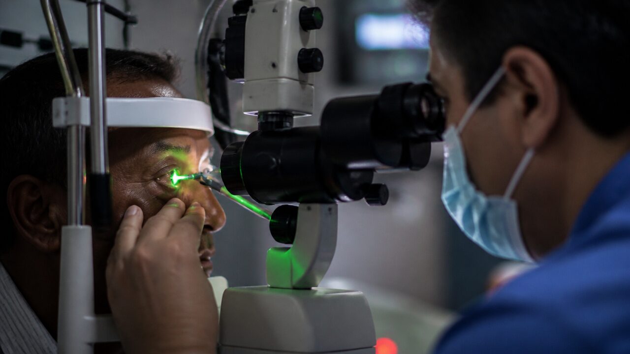
This treatment is carried out quite quickly and generally painlessly. The stages of the procedure are as follows. Preparation: drops are applied to numb your eye, so that you do not feel any pain during the laser treatment. Laser application: your doctor uses a special lens to direct the argon laser to the problem area in your eye. The laser repairs the vessels or tears that damage the retina. Healing: the treatment usually takes a few minutes, and afterwards there may be slight blurring in your eye. This blurring passes within a few hours.
Advantages of the Treatment
Protecting vision: argon laser photocoagulation can help prevent vision loss by treating retinal diseases early. Fast and painless: the procedure usually takes a short time, and because local anaesthesia is used no pain is felt. Minimally invasive: this method does not require surgery and allows patients to return to their daily lives quickly.
What to Expect After Treatment
After the laser treatment there may be blurring in vision and a mild feeling of discomfort for a few hours. This condition is usually short-lived, and most patients return to their normal vision levels within the same day. After the treatment, the following points should be noted: resting the eyes for a few days; using eye drops and medications regularly; and attending follow-up appointments as recommended by the doctor.
Are There Risks of Argon Laser Photocoagulation?
As with every medical procedure, argon laser photocoagulation can also have some risks. Although these risks are rare, the following situations may arise: temporary blurring or reduction in vision; problems in peripheral vision and in night vision; and the formation of scar tissue in the area where the procedure was performed. These risks can generally be kept under control when your doctor's advice is followed after the treatment.
This page is for general information and does not replace a personal examination. The right approach is decided together after an eye examination.