Glaucoma
Glaucoma is a progressive eye disease in which the optic nerve is damaged, often by raised eye pressure, and which can cause irreversible vision loss if not caught early.
What is glaucoma (eye pressure)?
Glaucoma is an eye disease that damages the optic nerve (the seeing nerve) and can lead to vision loss over time. It usually develops as a result of increased intraocular pressure, but in some cases optic nerve damage can occur even when the eye pressure is at a normal level. Essentially, the progressive deterioration of the eye nerve is what causes the disease. Glaucoma is one of the most common causes of permanent blindness worldwide and, if not detected early, can lead to permanent vision loss. It is an insidious disease that usually progresses without any obvious symptoms. For this reason it is also called the "silent thief of sight." Because vision loss is irreversible once it begins, regular eye examinations and early diagnosis are of great importance in preventing it.
What is the difference between glaucoma and eye pressure?
Glaucoma and eye pressure are two different concepts that are often confused with each other. Understanding the difference between these terms is important for diagnosing and treating glaucoma and for protecting eye health. Eye pressure can be one of the causes of glaucoma, but the two are not exactly the same thing.
Eye pressure refers to the intraocular pressure created by the fluid inside the eye (aqueous humor). This pressure helps the eye keep its shape and work correctly. Intraocular pressure is usually measured in mmHg (millimeters of mercury) and in a healthy eye should be between 10 and 21 mmHg. When the intraocular pressure is above this normal range, it is called high eye pressure. High eye pressure (ocular hypertension) does not by itself mean glaucoma unless it causes damage to the optic nerve. However, long-standing high intraocular pressure can damage the optic nerve, and in advanced stages this can turn into glaucoma. Eye pressure is a condition that can increase the risk of glaucoma, but not everyone with high eye pressure develops glaucoma. In other words, eye pressure generally refers to a rise in pressure inside the eye, while glaucoma is the name of the disease in which damage to the eye nerve has occurred.
Glaucoma is a vision-loss disease that occurs when the optic nerve in the eye is damaged because of high intraocular pressure or for other reasons. It is usually associated with raised intraocular pressure, but it can also develop in people whose eye pressure is normal. This shows that eye pressure can be a cause of glaucoma but is not its only cause. When glaucoma leads to vision loss, the damage it creates is irreversible, which is why early diagnosis is so important. Glaucoma is diagnosed when damage occurs in the optic nerve, even if the eye pressure is normal.
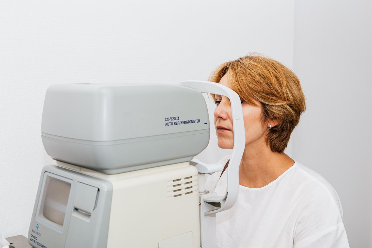
Eye pressure is measured by eye doctors using devices called tonometers. The tonometry test helps assess eye health by measuring the intraocular pressure. The test is painless and takes a few seconds. When high intraocular pressure is detected, close follow-up is needed and various measures are taken to prevent the development of glaucoma.
High eye pressure is a risk factor for glaucoma, but it is not sufficient on its own to make a diagnosis of glaucoma. Not everyone with high eye pressure develops glaucoma, but long-standing high intraocular pressure can damage the optic nerve and lead to glaucoma. In addition, in some people optic nerve damage (glaucoma) can develop even when the eye pressure is in the normal range. This condition is called normal-tension glaucoma.
In people with high eye pressure, treatment generally aims to lower the intraocular pressure. But if there is no evidence of any damage, this high pressure can simply be monitored, or, if it is thought to carry a risk of causing potential damage, pressure-lowering drops can be started. This is done to reduce the risk of developing glaucoma. Glaucoma treatment, on the other hand, aims to stop or slow optic nerve damage by bringing the intraocular pressure under control. Here, damage has already been proven and treatment is essential.
Early diagnosis is important for stopping the progression of glaucoma. Especially in people with high eye pressure, these risk factors should be followed and, if a risk of damage is seen, medication should be started. Do not neglect regular eye examinations if you have any of the following risk factors. Age: the risk of glaucoma increases in people over 40. Family history: the risk is higher if there is glaucoma in the family (especially in first-degree relatives). High intraocular pressure: people with high eye pressure carry a risk of developing glaucoma. Myopia: the risk is higher in people who are nearsighted. Diabetes and hypertension: health problems such as diabetes and high blood pressure can increase the risk of glaucoma. Long-term corticosteroid use: long-term use of such medications can raise the intraocular pressure.
Glaucoma and eye pressure are related but different conditions in terms of eye health. Although high eye pressure increases the risk of glaucoma, a diagnosis of glaucoma also requires optic nerve damage to be present. Keeping your intraocular pressure and optic nerve health under control with regular eye examinations is the most important step for the early detection and treatment of glaucoma. Especially people over 40, those with diabetes, and those with a family history of glaucoma should not neglect their regular eye checks.
What are the symptoms of glaucoma?
Glaucoma is an insidious disease that usually shows no obvious symptoms in the early stage. For this reason it is also called the "silent thief of sight." The symptoms of glaucoma can vary according to the type of disease and how quickly it progresses. Symptoms generally appear as the disease advances, and by this stage permanent vision loss may already have begun. Still, it can give some faint, vague signs.
The most common symptoms of glaucoma are as follows. Peripheral vision loss: glaucoma first usually affects side (peripheral) vision. The field of vision narrows over time and eventually only a central area may remain visible. Tunnel vision: as the disease progresses, the field of vision narrows and tunnel vision develops, leaving only a narrow central field of view; in this state patients cannot see objects around them and may bump into objects at the edges and have accidents at home or outside. Blurred vision: blurred vision can occur when the intraocular pressure rises or during an acute glaucoma attack. Light sensitivity: people with glaucoma may be more sensitive to bright lights and may see rings or colored halos around lights. Headache: a headache complaint can also occur, especially when the intraocular pressure is high. Eye pain: in some types of glaucoma, such as closed-angle glaucoma, there can be severe pain in the eye.
Open-angle glaucoma is the most common type of glaucoma and usually progresses slowly. This type may give no symptoms in the early stage. It is often detected incidentally during an eye examination. As it advances, the field of vision slowly narrows and a decrease in the eye's peripheral field of vision can be noticed.
Closed-angle glaucoma is a condition in which the intraocular pressure rises rapidly and which requires emergency intervention (an acute glaucoma attack). In this situation the symptoms are sudden and severe: severe eye pain that starts suddenly; headache, nausea and sometimes vomiting; blurred vision and loss of clear sight; redness in the white part of the eye with a sensation of hardening of the eye; and seeing colored rings around bright lights.
In normal-tension glaucoma, the intraocular pressure can be at normal levels but optic nerve damage still occurs. The symptoms can be similar to other types of glaucoma: peripheral vision loss, as in open-angle glaucoma, is the most common symptom, and central vision loss can appear in the advanced stage.
Congenital (present-at-birth) glaucoma is seen in babies and children and is a serious condition that needs to be diagnosed early. Symptoms can include constant watering of the eyes in infants, sensitivity to light with an inability to look at light and a desire to close the eyes, an abnormal enlargement of the affected eye, and cloudiness of the eye with opacity of the cornea.
Because glaucoma usually gives no symptoms in the early period, regular eye examinations are very important. Especially people over 40, those with a family history of glaucoma, and those with risk factors such as diabetes and hypertension should regularly have their intraocular pressure and optic nerve examined. With early diagnosis, the progression of glaucoma can be stopped and vision loss prevented. Remember: if you are experiencing one of the symptoms of glaucoma or are in the risk group for this disease, it is recommended that you see an eye doctor as soon as possible. When glaucoma is diagnosed early, it can be treated and vision loss can largely be prevented.
How do people with glaucoma see?
Glaucoma causes vision loss due to damage to the optic nerve that occurs as the intraocular pressure rises. This damage usually progresses slowly and the vision loss may not be noticed at first. People with glaucoma experience certain changes in their field of vision, especially as the disease advances. Depending on the type of glaucoma and how quickly it progresses, the signs of vision loss can also differ.
The most distinctive feature of glaucoma is that it usually causes vision loss starting from the peripheral (side) vision. Peripheral vision loss means that the outer parts of the field of view slowly disappear. As a result, people with glaucoma slowly lose their side vision (it may not be noticed at first, but later patients have difficulty seeing objects around them) and central vision may become the only remaining sight, so they can focus on the center but cannot see objects to the sides.
As glaucoma progresses and peripheral vision loss increases, patients can move into a state called tunnel vision. Tunnel vision is the narrowing of the field of view so that only a small central region can be seen clearly. Patients can clearly see only a narrow area in the center; because of peripheral vision loss the side parts appear completely dark or blurred. If the disease is not treated, the narrow remaining central area also gradually shrinks and patients feel as if they are looking through a tunnel.
In the final stages of glaucoma, the optic nerve is severely damaged. At this stage total vision loss can develop: the field of view becomes completely dark and blindness can occur. These advanced stages are irreversible: vision lost because of glaucoma cannot be regained, which is why early diagnosis and treatment are critically important.
The type of glaucoma can also affect how the patient sees. In open-angle glaucoma, the most common type, which usually progresses slowly, patients initially experience peripheral vision loss that goes unnoticed; over time side vision loss increases and tunnel vision occurs. In closed-angle glaucoma, which occurs with a sudden rise in intraocular pressure, patients experience symptoms such as sudden blurred vision, light halos and severe headache, and if not treated quickly it can lead to permanent vision loss. In normal-tension glaucoma the intraocular pressure is normal but optic nerve damage occurs; these people also gradually experience peripheral vision loss and progress toward tunnel vision.
A glaucoma patient's visual experience can be described like this. Healthy vision: normally people can see the surroundings over a wide angle and notice movements coming from the sides; the field of view is wide and central and peripheral vision are equally clear. Early-stage glaucoma vision: a slow narrowing of the field of vision begins and the side parts blur slightly. Advanced-stage glaucoma vision: the field of view is reduced to a narrow point in the center; patients can see clearly only a small central area while the surroundings are completely blurred or dark, which makes walking and daily tasks difficult. Because the vision loss experienced by glaucoma patients is irreversible, early diagnosis and treatment are very important. If you are experiencing any of the symptoms of glaucoma or are in the risk group, it is recommended that you see an eye doctor. Early diagnosis can stop vision loss and bring the progression of the disease under control.
What are the causes of glaucoma?
Glaucoma is an eye disease that usually develops as a result of increased intraocular pressure. The intraocular pressure rises when the fluids in the eye (especially the aqueous humor) cannot flow out or drain sufficiently. Since the eye is a closed sphere, an increase in fluid naturally leads to an increase in pressure, and the force of this pressure damages the eye nerve and over time leads to vision loss. However, the causes of glaucoma are not limited to intraocular pressure alone; many other factors can also increase the risk of glaucoma.
Blockage of intraocular fluid drainage: intraocular fluid leaves through special channels in the front of the eye called the trabecular meshwork. This fluid is continuously produced and drained to regulate the eye's internal pressure and maintain its structure. If these drainage channels become blocked or narrowed, fluid builds up in the eye and the intraocular pressure rises; this high pressure damages the optic nerve and leads to glaucoma.
Genetics and family history: genetic factors play an important role in glaucoma. If there is a family history of glaucoma, a person's risk of developing the disease increases. Glaucoma is associated with certain genetic mutations, and these genetic predispositions can raise the risk of the disease developing. Types such as primary open-angle glaucoma in particular are frequently seen in connection with genetic predisposition.
Age: aging is an important factor that increases the risk of glaucoma. Glaucoma is generally more common in people over 40. As age advances, the narrowing or loss of function of the channels that allow intraocular fluid to flow out can cause the intraocular pressure to rise.
High intraocular pressure: high intraocular pressure (ocular hypertension) is one of the most common risk factors for glaucoma. The intraocular pressure rises because the aqueous humor fluid continuously produced inside the eye cannot flow out. Intraocular pressure that remains high for a long time can damage the optic nerve and lead to the development of glaucoma.
Eye injuries and trauma: past eye injuries or trauma can increase the risk of glaucoma. Trauma to the eye can damage the drainage channels and cause the intraocular pressure to rise. This can lead immediately to the formation of glaucoma, or it can increase the risk of developing glaucoma even years later.
Long-term corticosteroid (cortisone) use: long-term use of medications containing corticosteroids can increase the intraocular pressure. The risk of glaucoma increases especially when eye drops or oral corticosteroid medications are used at high doses or for a long time. By blocking the drainage of aqueous humor in the eye, corticosteroids can raise the intraocular pressure and damage the optic nerve.
Diabetes and other health problems: health problems such as diabetes, hypertension and heart disease can increase the risk of glaucoma. Diabetes-related eye diseases such as diabetic retinopathy can raise the intraocular pressure and lead to glaucoma. Hypertension, by affecting the vessels in the eye, can reduce blood flow to the optic nerve and raise the risk of glaucoma.
Low eye pressure and blood-flow problems: in normal-tension glaucoma, the intraocular pressure can be at normal levels but blood-flow problems in the eye cause damage to the optic nerve. Low blood pressure and circulation problems can prevent the optic nerve cells from receiving enough oxygen and nutrients; in this case the optic nerve is damaged and glaucoma can develop.
Myopia and other eye-anatomy disorders: myopia (difficulty seeing far away) is one of the factors that increase the risk of glaucoma. In myopic eyes the internal anatomy is more prone to a rise in intraocular pressure. High-degree myopia in particular can put more pressure on the optic nerve. Other eye-anatomy disorders can also increase the risk of glaucoma.
Ethnic origin: ethnic origin is also a factor that affects the risk of glaucoma. For example, primary open-angle glaucoma is more common in African and African-American individuals and is more likely to appear at an early age. In people of Asian origin, the risk of closed-angle glaucoma is higher. These types of glaucoma can be seen more often and progress more severely depending on ethnic origin.
What should be done to prevent glaucoma?
Although it is not possible to prevent glaucoma with certainty, you can take some steps to reduce risk factors. Regular eye examinations: especially if you are over 40 or have risk factors for glaucoma, regular eye examinations are important; by having your intraocular pressure checked regularly you can monitor your risk of glaucoma. Healthy lifestyle: keeping in mind that health problems such as diabetes and hypertension increase the risk of glaucoma, adopt a healthy lifestyle: balanced nutrition, regular exercise and stress management can support your eye health. Avoiding eye injuries: eye injuries can increase the risk of glaucoma, so be careful to protect your eyes; using protective glasses, especially while doing sports or heavy work, can reduce the risk of injury. Following corticosteroid use: long-term corticosteroid use can increase the intraocular pressure, so use medications containing corticosteroids under a doctor's supervision and have your eye health checked regularly. Know your risk factors: if there is glaucoma in your family history, your risk may be higher, so monitor your eye health closely and have regular eye examinations before symptoms appear.
How is glaucoma diagnosed?
Because glaucoma is an important disease for eye health, early diagnosis is very critical. Various eye examinations and tests are used to diagnose glaucoma. These tests help determine whether glaucoma is present by assessing the intraocular pressure, the optic nerve and the field of vision. A diagnosis of glaucoma is made through a comprehensive examination performed by eye doctors. The main methods used when diagnosing glaucoma are described below.
Intraocular pressure measurement (tonometry): tonometry is a test used to measure the intraocular pressure. It is one of the most basic methods for assessing glaucoma risk. The test checks whether the intraocular pressure is above the normal range (generally 10–21 mmHg). High intraocular pressure increases the risk of glaucoma, but in some types of glaucoma (such as normal-tension glaucoma) the intraocular pressure can be at normal levels. The most commonly used method is applanation tonometry, in which the pressure is measured by lightly touching the surface of the eye; a local anesthetic drop is used to make the procedure painless. In air-puff (non-contact) tonometry, a simple test is performed by puffing air onto the surface of the eye; this method is fast and does not touch the eye surface.
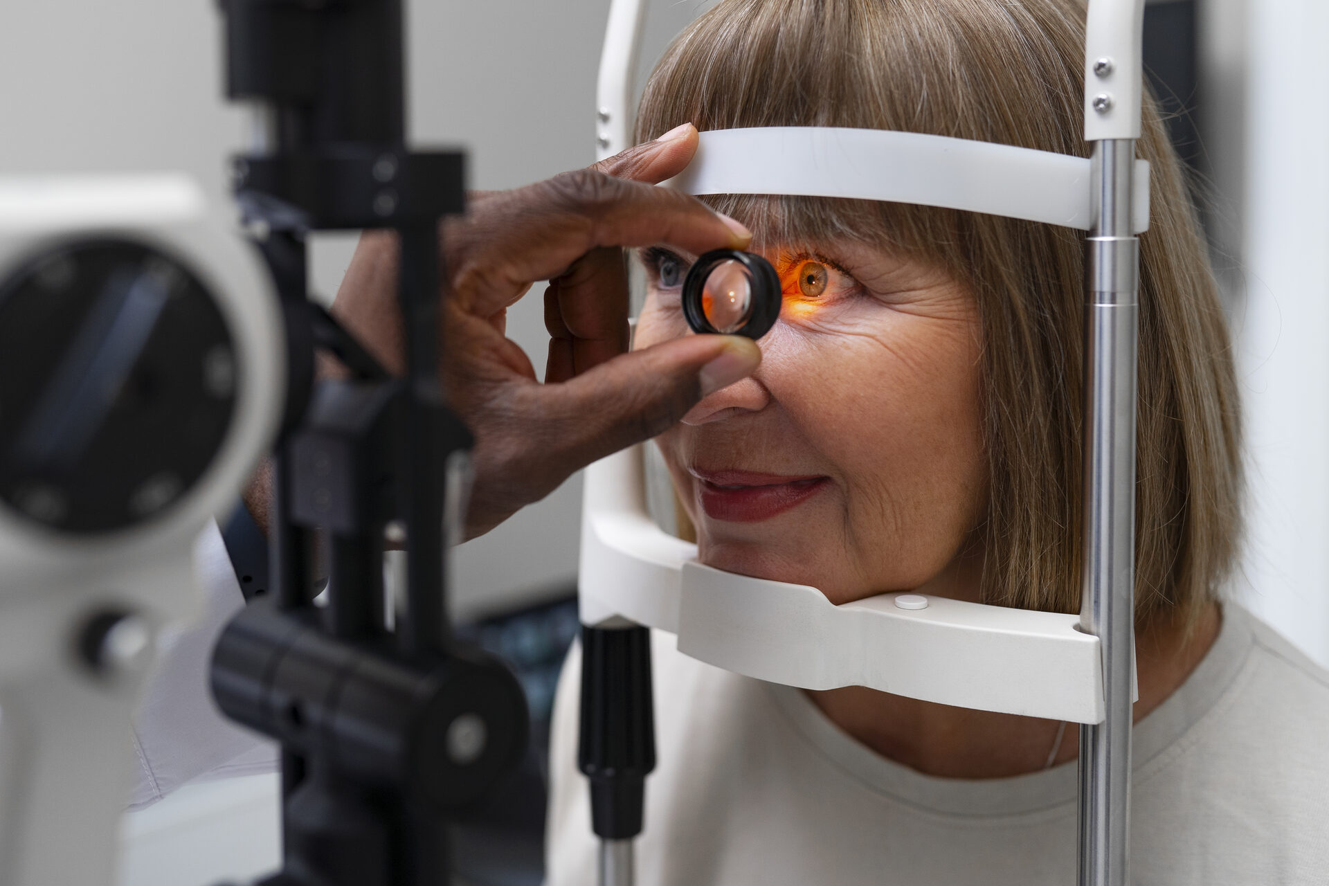
Optic nerve head (optic disc) examination (ophthalmoscopy): with a device called a biomicroscope, the optic nerve in the eye is viewed. The optic nerve is examined to assess whether there is glaucoma-related damage. Signs such as cupping (hollowing) or pallor of the optic nerve can point to glaucoma.
Visual field test (computerized perimetry): perimetry, or the visual field test, assesses the eye's peripheral vision (side vision). Because glaucoma usually causes loss of peripheral vision, this test can detect the areas affected by glaucoma. During the test the patient looks at a series of points of light and presses a button when they notice them; in this way a map of the eye's vision loss is produced. The visual field test is also performed regularly to monitor the progression of glaucoma. The degree of vision loss provides important information for understanding what stage the disease is at.
Gonioscopy: gonioscopy allows the eye's drainage angle to be assessed. Glaucoma is classified as open-angle or closed-angle, and this test helps determine the type of glaucoma. By examining the trabecular meshwork and the drainage angle, the doctor checks how the intraocular fluid is being drained. In open-angle glaucoma the outflow path for the intraocular fluid is open but the drainage is slow; in closed-angle glaucoma the outflow path for the fluid is closed and it is a condition requiring emergency intervention.
Optical coherence tomography (OCT): OCT is a test that provides detailed images of the retina and the optic nerve fibers. It is used to image glaucoma-related thinning and damage in the optic nerve and retinal layer. This method measures the detailed structure and thickness of the optic nerve. OCT helps diagnose glaucoma at an early stage and monitor the progression of the disease.
Pachymetry (corneal thickness measurement): pachymetry is a test that measures the thickness of the cornea. Corneal thickness can affect intraocular pressure measurements. For example, the risk of developing glaucoma is higher in people with thin corneas. These results help in assessing the intraocular pressure more accurately. This test is an important complementary method in the diagnosis of glaucoma.
Because glaucoma usually progresses without giving early symptoms, it is important, especially for people in the risk group, to have regular eye examinations. Early diagnosis is critically important for slowing the progression of glaucoma and preventing vision loss. If you are over 40, have a family history of glaucoma, or have health problems such as diabetes or high blood pressure, you can manage your glaucoma risk by having regular eye examinations. When diagnosing glaucoma, several tests are usually performed together (tonometry, ophthalmoscopy, perimetry, gonioscopy and OCT) so that the doctor can assess the intraocular pressure, the optic nerve and the field of vision in detail. Early diagnosis is of great importance for stopping or slowing the progression of glaucoma.
Is glaucoma genetic?
Yes, glaucoma has a genetic component, and the likelihood of seeing this disease is higher in people with a family history of glaucoma. Genetic factors are one of the important elements that increase the risk of glaucoma. Glaucoma can arise through the effect of more than one gene, and sometimes it can develop because of a mutation in a single gene. However, environmental factors, age and other health problems can also play a role in the appearance of glaucoma.
Genetic predisposition can differ among the various types of glaucoma. In primary open-angle glaucoma, the most common type, genetic factors play an important role; if there is a family history of primary open-angle glaucoma, individuals are more likely to develop this type. Mutations in genes such as MYOC, OPTN and WDR36 are known to increase the risk of primary open-angle glaucoma; these genes play a role in producing proteins that regulate intraocular pressure. Closed-angle glaucoma can also be influenced by genetic predisposition; it is more common in people of Asian origin, and the risk is higher in those with a family history. The anatomical structure of the eye is thought to be inherited, and the risk of closed-angle glaucoma may be higher in individuals with a narrow anterior chamber angle. Congenital glaucoma is one of the types in which genetic factors are most prominent; it usually appears in babies or young children and can be inherited from the family: mutations in genes such as CYP1B1 and LTBP2 have been associated with it and can adversely affect the development of the eye's fluid drainage channels. In normal-tension glaucoma, genetic factors can also play a role even though the intraocular pressure is at normal levels, and additional risk factors such as low blood pressure, migraine and vascular irregularities are present alongside genetic predisposition.
The risk of developing glaucoma is higher in people with a family history of the disease. The genetic components of glaucoma can affect the mechanisms that regulate intraocular pressure and the resilience of the optic nerve. In people with a genetic predisposition there can be genetic mutations related to the drainage of intraocular fluid, causing the fluid to be unable to drain and the pressure to rise; the resilience of the optic nerve is related to genetic factors, so since glaucoma causes optic nerve damage, genetic predisposition can affect the nerve's capacity to withstand this pressure; and eye anatomy can be inherited within a family: the risk of closed-angle glaucoma in particular is directly related to eye anatomy and is more common in people with a narrow-angle eye structure.
If there is a family history of glaucoma, having your eye health checked regularly is of great importance. Because people with glaucoma in the family are known to carry a higher risk, assessments such as intraocular pressure measurement and optic nerve examination should be done regularly. Early diagnosis plays a critical role in preventing the vision loss caused by glaucoma in people with a genetic predisposition. In some cases, genetic tests can be used to obtain information about genetic predisposition, especially in certain types such as congenital glaucoma. However, in most types of glaucoma, genetic tests are not routinely applied. It is more important for people with a family history of glaucoma to know their genetic predisposition, have regular eye examinations and manage their risks.
Glaucoma can develop through the effect of environmental and lifestyle factors together with genetic factors. While genetic predisposition forms a basis, environmental factors can accelerate the onset of the disease. These factors can include aging (eye health can change with age and the risk of glaucoma increases), diabetes and hypertension (these diseases can increase glaucoma risk by affecting blood flow inside the eye), and long-term corticosteroid use (corticosteroid-containing medications can raise the intraocular pressure and increase the risk of glaucoma). Although genetic predisposition is a factor that increases the risk of glaucoma, it is possible to detect and manage glaucoma early through a healthy lifestyle and regular eye examinations. Therefore, it is of great importance for individuals with a family history of glaucoma to have regular eye examinations and to have their intraocular pressure and optic nerve checked. Regular follow-up and early diagnosis are the most effective way to stop the progression of glaucoma and prevent vision loss.
Is glaucoma a temporary disease? Does glaucoma go away completely?
No, glaucoma is not a temporary disease. It is usually a chronic and progressive eye disease. It develops as the optic nerve is damaged by a rise in intraocular pressure and can lead to permanent vision loss. When glaucoma is not treated, the damage to the optic nerve is irreversible. For this reason, it is very important for glaucoma to be diagnosed and treated early. The optic nerve damage caused by glaucoma is permanent, and as it progresses the vision loss is irreversible. However, with regular treatment and follow-up, glaucoma can be brought under control so that the progression of the disease can be stopped or slowed. By lowering the intraocular pressure with treatment, the pressure on the optic nerve is reduced and further vision loss is prevented.
Because glaucoma is a progressive disease, it requires regular treatment and follow-up. With glaucoma treatment the progression of the disease can be stopped, but the existing damage is irreversible. Treatment options include eye drops, which are the most common treatment method: pressure-lowering drops used regularly reduce the pressure on the optic nerve; laser treatment, which can be applied to improve the outflow of fluid in the eye and help lower the intraocular pressure; and surgical intervention, which in advanced stages can be used to increase the drainage of intraocular fluid, with these operations trying to prevent optic nerve damage by lowering the pressure. Untreated glaucoma can over time lead to vision loss and ultimately permanent blindness. Because glaucoma gives no symptoms in the early period, patients usually notice it late, which is why regular eye examinations are very important. Glaucoma detected early can be brought under control with treatment and further vision loss prevented.
What is good for glaucoma?
Glaucoma is a chronic eye disease that needs to be treated. Although it is not possible for glaucoma to heal completely, it is possible to bring the progression of the disease under control by lowering the intraocular pressure. This can be achieved with regular treatment, healthy lifestyle changes and adherence to your eye doctor's advice. The main treatment methods are summarized below, together with some lifestyle recommendations.
Eye drops are the most common treatment method for glaucoma. These drops reduce the pressure on the optic nerve by lowering the intraocular pressure. The drops used in glaucoma treatment generally include prostaglandin analogues (which lower the pressure by increasing the outflow of fluid in the eye), beta blockers (which reduce the production of intraocular fluid), and alpha agonists and carbonic anhydrase inhibitors (which lower the intraocular pressure both by reducing fluid production and by increasing drainage). Eye drops should be used regularly as recommended by the doctor; interrupting or misusing the treatment can lead to the intraocular pressure not being controlled.
Laser treatment can be used to improve the outflow of fluid in the eye and can be an effective solution for lowering the intraocular pressure in glaucoma patients. Laser trabeculoplasty increases fluid flow by opening the drainage channels in the eye and is generally used in open-angle glaucoma patients. Laser iridectomy is used in closed-angle glaucoma patients to open a blockage in the iris layer.
Surgical intervention may be needed for glaucoma patients in advanced stages. Surgical treatments aim to lower the intraocular pressure by creating new paths that allow fluid to drain out of the eye. These operations can include trabeculectomy (a new drainage path is opened for the fluid to leave the eye), minimally invasive glaucoma surgeries, and shunt placement (a tube is placed to help drain fluid out of the eye).
Although various supportive measures have shown benefit in some studies on glaucoma, there is still no supportive measure that we recommend to our patients and say "you must also do this." But I believe that everything we can do for a healthy life may also benefit chronic diseases. For this reason my recommendations are as follows. Regular exercise: regular moderate-intensity exercise can help lower the intraocular pressure; however, some exercises (especially head-down positions) can raise it, so you should discuss your exercise plan with your doctor. Healthy nutrition: a balanced diet is important for supporting eye health, and it can be beneficial to consume plenty of antioxidant-rich vegetables (such as green leafy vegetables), fish rich in omega-3 fatty acids, and vitamins such as C and E that protect eye health. Stress management: stress can raise the intraocular pressure, and stress-management techniques such as yoga, meditation and breathing exercises can indirectly improve your eye health. Limit smoking and alcohol: smoking and excessive alcohol use can harm eye health and increase the risk of glaucoma. Limiting caffeine: excessive caffeine intake can temporarily raise the intraocular pressure in some people, so if you have glaucoma it can be helpful to avoid excessive coffee and tea.
Do not neglect doctor checks: glaucoma is a disease that requires regular follow-up; it is very important to have your intraocular pressure measured and your optic nerve health monitored at the intervals set by your eye doctor. Because glaucoma is a progressive disease, its progression can be slowed or stopped with regular checks. Pay attention to your sleeping position: sleeping face-down or lying on one eye can raise the intraocular pressure in some people; keeping your head slightly elevated while sleeping can reduce the pressure, and choosing a suitable pillow can help control it. It should also be kept in mind that hypotensive episodes during sleep can increase damage by reducing blood supply to the eye nerve, so precautions against this should be taken.
Some natural treatments can be used to support eye health, and their benefit has been shown in some studies. However, these absolutely do not replace treatment; they can be supportive in addition to the treatment recommended by your doctor. Omega-3 fatty acids, which are good for eye health, are found especially in fish; these acids can protect the retina and reduce inflammation in the eye. Ginkgo biloba can help increase blood flow and improve blood circulation in the eyes, but you should consult your doctor before using it. Green tea, which is rich in antioxidants, can protect eye health, but it should be used carefully because excessive consumption can raise the intraocular pressure. Although glaucoma cannot be completely cured, it can be brought under control with treatments that lower the intraocular pressure and with lifestyle changes. Carefully following your eye doctor's recommendations and having regular checks are very important in glaucoma treatment.
What are the types of glaucoma?
Glaucoma is a group of eye diseases in which the optic nerve is damaged as a result of increased intraocular pressure. Glaucoma is divided into different types, and the development mechanism, symptoms and treatment approaches of each type can differ.
Primary open-angle glaucoma is the most common type of glaucoma and usually progresses slowly. Because the intraocular fluid (aqueous humor) cannot be drained out of the eye quickly enough, the intraocular pressure slowly rises. This type usually gives no symptoms, and by the time vision loss is noticed the disease has often reached an advanced stage; peripheral (side) vision loss occurs first. Factors such as family history, advanced age, myopia, diabetes and high blood pressure increase the risk. Treatment tries to lower the intraocular pressure using eye drops, laser treatment or surgical methods.
Closed-angle glaucoma (angle-closure glaucoma) occurs as a result of the sudden narrowing or closure of the drainage angle in the eye (the region in the front of the eye that allows fluid to drain out). The intraocular pressure rises rapidly and can cause sudden damage to the optic nerve; this is called acute angle-closure glaucoma. It shows itself with symptoms such as severe eye pain, headache, blurred vision, redness of the eye, nausea and seeing halos around lights, and it requires emergency medical intervention. It is more common in people of Asian origin and in individuals with narrow eye angles. Because closed-angle glaucoma is an emergency, rapid treatment is required: laser iridectomy is the procedure of opening a small hole in the iris to allow the flow of intraocular fluid, and eye drops and sometimes surgical treatment may also be needed.
Normal-tension glaucoma (low-tension glaucoma) develops while the intraocular pressure is normal. In this type, even though the intraocular pressure is in the normal range, damage occurs in the optic nerve. The reason for this is not fully understood, but factors such as impaired blood circulation in the eye or the optic nerve being more sensitive to normal pressure may be responsible. Peripheral vision loss and, in the advanced period, central vision loss can be experienced. It may be more common in people with circulation problems such as migraine, low blood pressure and cold hands and feet. Eye drops, laser treatment or surgical methods are used to lower the intraocular pressure even further.
Secondary glaucoma arises from another condition: another disease, injury, inflammation or medication use. For example, conditions such as uveitis (inflammation in the eye), eye injuries and long-term corticosteroid use can lead to secondary glaucoma. The symptoms can vary depending on the underlying disease or condition. Treatment aims to remove the underlying cause and bring the intraocular pressure under control; eye drops, laser treatment or surgical methods can be used.
Congenital glaucoma (glaucoma present at birth) is seen in babies and children. It is a rare type of glaucoma that can be seen in babies from birth and usually develops as a result of the eye's drainage channels not developing or not working sufficiently from birth. In babies there can be excessive watering of the eye, excessive sensitivity to light, an increase in eye size (buphthalmos) and cloudiness of the eye. Treatment of congenital glaucoma is usually surgical; surgical interventions are performed to open the drainage paths and bring the intraocular pressure under control.
Pigmentary glaucoma arises from pigment cells. It occurs when the pigments in the iris layer of the eye block the eye's drainage system, and this blockage can cause the intraocular pressure to rise. Symptoms can include vision loss, blurred vision and seeing halos around lights. The intraocular pressure can be brought under control with eye drops, laser treatment and surgical methods.
Exfoliative glaucoma (pseudoexfoliation syndrome) arises from deposits on the iris and lens. It occurs when fine, dandruff-like material that accumulates on the iris and lens in the eye blocks the drainage system. This type of glaucoma, like open-angle glaucoma, usually progresses slowly and gives no symptoms, and vision loss occurs over time. Eye drops, laser treatment and surgical methods can be applied. Glaucoma has different types, and the development mechanism and treatment approach of each are different. If glaucoma is not detected early it can lead to permanent vision loss, so regular eye examinations are very important: especially if you are in the risk group (family history of glaucoma, diabetes, high blood pressure, advanced age). Your doctor will create the appropriate treatment plan according to the type of glaucoma.
What is an acute glaucoma attack?
An acute glaucoma attack is a type of glaucoma that appears suddenly and severely. It occurs as a result of a sudden and rapid rise in intraocular pressure and shows itself with symptoms such as severe pain in the eye and vision loss. It occurs in the type known as closed-angle glaucoma and requires emergency medical intervention. If not treated quickly, optic nerve damage and permanent blindness can develop.
An acute glaucoma attack develops as a result of the sudden closure or narrowing of the drainage channels for the fluid inside the eye. Normally the fluid produced in the eye (aqueous humor) is drained out through a channel called the trabecular meshwork. However, in closed-angle glaucoma this drainage angle closes and the intraocular fluid cannot flow out, which causes the intraocular pressure to rise rapidly. Closed-angle glaucoma is usually seen in people with an anatomical predisposition, and the risk is greater especially in individuals with a narrow anterior chamber angle. Risk factors include age (it is more common in people over 40), sex (it may be more common in women), ethnic origin (the risk is higher in people of Asian origin) and family history (the risk increases in individuals with a family history of glaucoma).
An acute glaucoma attack shows itself with sudden and severe symptoms that occur as a result of the rapid rise in intraocular pressure. Severe eye pain: a very severe pain that starts suddenly is felt in the eye, and it can sometimes spread to the face and head region. Blurred vision: as the intraocular pressure rises, vision blurs and vision loss occurs. Headache: the high pressure in the eye can cause a severe headache, usually concentrated around the forehead and eyes. Nausea and vomiting: the severe headache and eye pain are usually accompanied by nausea and vomiting. Seeing halos around lights: patients may see colored rings around light sources. Redness of the eye: there can be a marked redness and watering of the eye. Hardening of the eye: with the rise in intraocular pressure, a hardening and tension of the eye is felt. Because an acute glaucoma attack progresses very quickly, an eye doctor must be seen urgently as soon as these symptoms appear; if not treated, the optic nerve can be damaged and permanent blindness can develop.
Because an acute glaucoma attack is an emergency, it must be treated immediately. Treatment aims to rapidly lower the intraocular pressure and open the drainage angle. The first treatment in an acute glaucoma attack is usually with medication: eye drops, oral medications and intravenous medications (mannitol given through a vein) can be used to lower the intraocular pressure rapidly, working by reducing fluid production in the eye or increasing the outflow of fluid. After the intraocular pressure is brought under control with medication, laser treatment can be applied. The laser treatments frequently used in an acute glaucoma attack are laser iridectomy (a small hole is opened in the iris layer to allow the outflow of intraocular fluid, reopening the drainage angle and lowering the pressure) and laser trabeculoplasty (performed to open the drainage channels and allow the intraocular fluid to flow better). If medications and laser treatment are not sufficient, surgical intervention may be needed; surgical methods open paths that allow the fluid to drain out and aim to bring the intraocular pressure under control.
An acute glaucoma attack is a very rapidly developing condition that can lead to permanent vision loss. The intraocular pressure can cause serious damage to the optic nerve in a short time, so an eye doctor must be seen as soon as the symptoms are noticed; with early intervention the risk of optic nerve damage and blindness can be minimized. If you are known to have a narrow-angle eye anatomy or carry a risk of closed-angle glaucoma, preventive measures can be taken to protect against an attack. Regular eye examinations let you have your intraocular pressure and drainage angle checked. If your doctor thinks your eye structure is prone to an acute glaucoma attack, a preventive laser iridectomy can be performed before an attack develops. Using the eye drops your doctor prescribes regularly to keep the intraocular pressure under control can reduce the risk of an attack. An acute glaucoma attack is an emergency in which closed-angle glaucoma appears suddenly and severely and can cause permanent damage in the eye; the rapid rise in intraocular pressure can damage the optic nerve and, if untreated, permanent blindness can develop within days, so when an acute glaucoma attack is experienced an eye doctor must be seen urgently and the necessary treatment applied.
What is the computerized visual field test?
The computerized visual field test (perimetry) is a test that measures the eye's peripheral vision ability and assesses losses in the field of vision. This test is used especially in the diagnosis and follow-up of diseases such as glaucoma. The computerized visual field test assesses eye health by detecting whether there is any narrowing, gap or abnormality in the field of vision. The concept we call the visual field refers to the entire peripheral area a person can see while looking steadily at one point. Under normal conditions not only the point looked at directly but also the objects around that point are seen within a certain boundary. The visual field test measures this breadth and sensitivity.
The computerized visual field test is usually carried out as follows. Preparation: the patient sits in front of a computerized device and the chin is fixed on a support; one of the eyes is covered while the other is asked to focus directly on a point, and this fixed point must be looked at during the test. Responding to points of light: the device sends points of light to different places in the field of vision, and the patient informs the device by pressing a button in their hand the moment they see these lights; in this way it is measured which points in the eye's peripheral area it can perceive. Visual field map: at the end of the test the device maps the points of light the eye perceived and the areas in which the eye shows weakness; this map helps the eye doctor understand the location and degree of vision loss. The test usually takes 10–20 minutes and is painless; the results are obtained immediately and assessed by the doctor.
The computerized visual field test holds a very important place especially in the early diagnosis and follow-up of diseases such as glaucoma, and it is also applied for many eye and nerve diseases. In glaucoma the first visual field to be lost is usually the side (peripheral) field of vision, and the test is used in diagnosis and in following progression; it is important for glaucoma patients to have this test at regular intervals to control the progression of the disease. Retinal diseases can also cause narrowing of the visual field, and the test helps in diagnosing conditions such as retinal degenerations and retinal tears. Diseases of the optic nerve (such as optic neuropathy) can cause losses in the field of vision, and the test is used to understand whether there is damage to the optic nerve. Tumors located in the brain or the optic nerve and other neurological diseases can lead to losses in the field of vision, and the test is also used to assess the effects of central nervous system diseases on the eye. Patients who have had a stroke or head trauma can also have visual field losses, and the computerized visual field test is performed to assess such damage.
The results of the visual field test are carefully assessed by the eye doctor. The results map the eye's peripheral vision ability, and this map shows whether there are gaps or weaknesses in the field of vision. If the test results are normal, this shows that the eye has a wide field of vision and there is no loss in peripheral vision. If vision loss, narrowing or gaps in certain regions are detected, this can point to glaucoma, retinal diseases or optic nerve diseases, and such situations may require the eye doctor to perform more detailed examinations. Blind spots called scotomas can also be detected in the field of vision; these blind spots show that there is vision loss in certain areas of the eye.
The computerized visual field test is critically important especially for the early diagnosis of insidiously progressing diseases that initially give no symptoms, such as glaucoma. In glaucoma, while central vision is preserved, side vision is slowly lost; this may not be noticed, but these losses can be detected with the visual field test. The test is used not only for diagnosis but also to assess the effectiveness of glaucoma treatment; the progression of the disease can be monitored with regular visual field tests and treatment can be adjusted according to these results. The computerized visual field test is an important test used in assessing eye health and plays a critical role especially in the early diagnosis of eye diseases such as glaucoma; it detects peripheral vision losses and is used in the diagnosis and monitoring of conditions such as glaucoma, retinal diseases, optic nerve damage, neurological diseases and stroke.
The computerized visual field test in glaucoma
Glaucoma is an eye disease that especially causes peripheral (side) vision loss. One of the most important tests in diagnosing glaucoma and monitoring its progression is the computerized visual field test. By determining the degree of vision loss in the eye and in which regions it occurs, this test helps in diagnosing glaucoma and in monitoring the response to treatment. Because glaucoma is an insidiously progressing disease, it usually gives no obvious symptom in the early period. However, losses in the visual field can begin in the first stages of the disease. In glaucoma patients, peripheral (side) vision is affected first and, in the advanced period, leads to the condition called tunnel vision; the computerized visual field test is used to detect these losses in the early period.
The computerized visual field test in glaucoma is critically important for several reasons. Early diagnosis of glaucoma: in glaucoma the first field of vision to be lost is usually peripheral vision, and patients may not notice this loss because central vision can be preserved for a long time; the test detects such peripheral vision losses in the early stages. Monitoring the progression of glaucoma: in glaucoma treatment, pressure-lowering medications or surgical interventions are used, and the test is performed to monitor the effectiveness of these treatments; tests done at regular intervals show whether glaucoma is progressing and how successful the treatment is. Creating a visual field map: the test detects whether there are any gaps in the patient's field of vision, and these gaps show that the optic nerve in the eye is damaged and that vision loss is occurring in that region; as glaucoma progresses these gaps widen, and this process can be monitored through the test.
The computerized visual field test is repeated at certain intervals in glaucoma patients and the rate of progression of the disease is monitored. In the test preparation the patient sits in front of a computerized device with the head fixed on a chin rest; one eye is covered while the other is asked to look directly at a point. The device sends points of light of different sizes and brightnesses to different areas of the eye, and the patient responds by pressing a button to each point of light they see. The device records which points the patient saw and which they could not see, and these results are assessed by the eye doctor to determine whether there is loss in the field of vision. At the end of the test a visual field map is created showing in which regions of the eye there is vision loss and the degree of this loss; in glaucoma patients this map can show how far the optic nerve damage has progressed.
The results of the visual field test in glaucoma patients are carefully assessed by the eye doctor and help in understanding what stage the disease is at and the degree of damage in the optic nerve. A normal visual field result means there is not yet an obvious vision loss in the eye, but this does not mean there is no glaucoma, so it must be assessed together with the other glaucoma tests. In glaucoma the first field of vision to be lost is usually the peripheral field, so if there is loss in the side field this can be an early sign of glaucoma. Blind spots called scotomas can be detected; scotomas show the regions with vision loss due to optic nerve damage and usually widen over time in glaucoma patients. As glaucoma progresses the losses in the field of vision increase, and with regularly performed tests these losses can be monitored and the response to treatment assessed.
The visual field test in glaucoma is of vital importance for understanding the rate of progression of the disease and monitoring the effectiveness of treatment. Because glaucoma causes permanent damage in the optic nerve, regularly performed visual field tests are used to track how far this damage has progressed. The benefits of the regular visual field test include early intervention (detecting loss before it begins so the treatment plan can be arranged), monitoring adherence to treatment (the effect of eye drops or surgical interventions can be assessed) and following disease progression (how quickly glaucoma is progressing and the general state of eye health can be seen). The computerized visual field test is one of the most important tests in the diagnosis and follow-up of glaucoma patients; in the early periods of glaucoma side (peripheral) vision loss usually begins, and this loss can be detected with the visual field test. The test should be performed regularly both for diagnosis and to assess the effectiveness of glaucoma treatment. If you have glaucoma or are at risk, it is very important to have visual field tests at the intervals determined by your eye doctor; these tests help to monitor the progression of glaucoma and minimize vision loss.
Eye tomography (OCT) in glaucoma
OCT (optical coherence tomography) is an advanced imaging method used in the diagnosis and follow-up of eye diseases such as glaucoma. By imaging detailed cross-sections of the retina and the optic nerve located at the back of the eye, OCT allows these structures to be examined. Because glaucoma damages the optic nerve, OCT is a very valuable test for detecting how much this nerve is affected and the extent of the damage. The use of OCT to diagnose the optic nerve damage caused by glaucoma early and to monitor the effectiveness of treatment has become widespread; your eye doctor being able to examine the state of your eye in detail with OCT can make a very big difference in planning treatment.
OCT is a non-invasive test (one that does not require intervention inside the eye) that produces high-resolution images of the various layers of the eye. By imaging the tissue layers in the eye at the micrometer level, it detects fine changes in the structure of the optic nerve and retina. The OCT test is painless and is usually completed within a few minutes. The image is taken without any contact with your eye, just like taking a photograph. The eye doctor may use a drop to enlarge your pupil before the OCT, which allows clearer images to be obtained. OCT creates detailed cross-sections of the retina and optic nerve in your eye, and these cross-sections are assessed by the eye doctor; the optic nerve fiber thickness in particular is an important criterion for monitoring the progression of glaucoma.
In glaucoma patients, OCT is used to detect the extent of thinning or damage in the optic nerve fibers. As glaucoma progresses the optic nerve fibers thin, and through OCT this thinning can be monitored precisely. OCT plays several key roles in glaucoma management. Measuring optic nerve fiber thickness: one of the most critical points in glaucoma is damage to the optic nerve fibers; OCT reveals how far the damage in the optic nerve has progressed by measuring the thickness of these fibers, which is an important criterion for monitoring the success of treatment: a healthy optic nerve is normally thick and sound, while a glaucomatous optic nerve thins as it progresses, causing vision loss. Monitoring the retinal ganglion cells: OCT can also image the retinal ganglion cells, which are a part of the optic nerve fibers that carry visual signals to the brain; because these cells are also affected by glaucoma, the state of the retinal ganglion cell layer is assessed with OCT. Early diagnosis of glaucoma: in the early stages of glaucoma obvious vision loss may not be noticed, but OCT can detect the damage in the optic nerve before vision loss begins, so that treatment can be started earlier and the damage brought under control. Assessing the response to treatment: OCT can be done regularly during the glaucoma treatment process so that changes in the optic nerve fibers can be followed, and the results are of great importance for monitoring whether the treatment is effective and updating the treatment plan if necessary.
The advantages of OCT include that it is non-invasive (a safe test applied without intervention inside the eye), allows early diagnosis (it can detect the optic nerve damage caused by glaucoma at very early stages), provides detailed images (very detailed images of the retina and optic nerve are obtained) and supports treatment follow-up (it is a reliable method for monitoring the effect of treatment). When glaucoma is diagnosed and the treatment process is started, the OCT test should be done at regular intervals; your doctor will decide how often you need to have this test depending on the rate of progression of glaucoma and your response to treatment. Generally, glaucoma follow-up with OCT is done once or twice a year. OCT is a highly effective and reliable method in the treatment and follow-up of glaucoma; by detecting changes in the optic nerve early, it is very important for optimizing your treatment plan and stopping vision loss. Regular OCT follow-up in the fight against glaucoma will provide a great advantage in protecting your eye health.
Is glaucoma dangerous?
Yes, glaucoma is a dangerous eye disease and, if not treated, can lead to permanent vision loss and even blindness. Glaucoma is a progressive disease that damages the optic nerve in the eye. The optic nerve is the structure that carries visual signals from the eye to the brain, so damage to this nerve causes the loss of sight. The most dangerous aspect of glaucoma is that it usually gives no symptoms in the early stages of the disease and progresses insidiously. Many people may begin to experience permanent vision loss because of glaucoma without realizing it, which is why early diagnosis and regular treatment are of vital importance.
Glaucoma causes damage to the optic nerve, and this damage is permanent. Optic nerve cells cannot be regenerated, so the vision loss caused by glaucoma is irreversible. In the advanced stages of glaucoma, patients begin to experience peripheral vision loss (side vision loss), and this loss may not be noticed; over time central vision is also affected and ultimately blindness can develop. The biggest danger of glaucoma is that it gives no symptoms in the early stages; many people may already have experienced vision loss by the time they realize they have glaucoma. Because glaucoma usually progresses slowly and peripheral vision loss is not noticed, patients can realize the disease late, so it is of vital importance for people at risk to have regular eye examinations. Some types of glaucoma, especially closed-angle glaucoma, can appear suddenly; during an acute glaucoma attack the intraocular pressure rises rapidly and shows itself with symptoms such as severe eye pain, blurred vision, headache and nausea, and this attack requires emergency intervention: if not treated it can cause permanent vision loss and blindness. The risk of glaucoma increases with age and is more common in people over 40; in addition, the likelihood of developing glaucoma is higher in those with risk factors such as diabetes, high blood pressure and a family history of glaucoma, so it is important for these people to have regular eye examinations and to have their intraocular pressure checked.
Although glaucoma is a dangerous disease, vision loss can be stopped or slowed with early diagnosis and treatment. The early diagnosis of glaucoma is very important for preventing the permanent vision loss it causes; especially people over 40 and those with risk factors for glaucoma should have regular eye examinations, and glaucoma can be diagnosed with tests such as intraocular pressure measurement (tonometry), optic nerve examination and visual field tests. When glaucoma is diagnosed, the treatment recommended by the doctor must be followed strictly; the intraocular pressure can be brought under control with eye drops, laser treatment or surgical interventions, and treatment helps to stop or slow the progression of glaucoma: if treatment is interrupted the damage in the optic nerve continues and more vision loss can occur. Bringing risk factors such as diabetes and high blood pressure under control can slow the progression of the disease, and adopting a healthy lifestyle, exercising regularly and eating healthily can reduce the risk of glaucoma. Glaucoma can be linked to genetic predisposition; the risk of the disease is higher in people with a family history, so if family members have a history of glaucoma you can reduce risk by having regular eye examinations.
Some types of glaucoma can progress more quickly and dangerously. Open-angle glaucoma is the most common type and usually progresses slowly, but if not detected early it can lead to serious vision loss. Closed-angle glaucoma shows itself with a sudden rise in intraocular pressure, requires emergency intervention and can cause sudden vision loss. Normal-tension glaucoma involves optic nerve damage even though the intraocular pressure is at normal levels; this potentially dangerous type can be detected with regular eye examination. When untreated, glaucoma is a dangerous disease that can lead to permanent blindness. Its most dangerous aspect is that it gives no symptoms in the early stages and progresses insidiously. Because the vision loss caused by glaucoma is irreversible, early diagnosis and treatment are critically important. Having regular eye examinations, having your intraocular pressure checked and being careful about glaucoma risk factors are the most effective ways to bring this dangerous disease under control.
Glaucoma treatment
Glaucoma is an eye disease that progresses by damaging the optic nerve and, when untreated, can result in permanent blindness. The main aim in glaucoma treatment is to prevent or slow optic nerve damage by bringing the intraocular pressure under control. Several different methods are used in glaucoma treatment, and the treatment plan is determined depending on the patient's condition, the type of glaucoma and the degree of progression of the disease. Treatment methods include eye drops, medication, laser treatment and surgical interventions. The aim of glaucoma treatment is to prevent further damage to the optic nerve and to lower the intraocular pressure, because the vision loss caused by glaucoma is irreversible.
The most common treatment method for glaucoma is eye drops. These drops reduce the pressure on the optic nerve by lowering the intraocular pressure and prevent further damage. The eye drops used in glaucoma treatment have several different mechanisms of action. Prostaglandin analogues lower the intraocular pressure by increasing the outflow of fluid in the eye; they are generally used once a day, and side effects such as redness of the eye, lengthening of the eyelashes and a change in eye color can be seen. Beta blockers lower the intraocular pressure by reducing the amount of fluid produced in the eye; they are generally used once or twice a day, and because they can have effects on heart rhythm and blood pressure they should be used carefully in patients with cardiovascular disease. Alpha agonists both reduce intraocular fluid production and increase its outflow; they can be applied several times a day, and side effects such as dryness of the eye, dry mouth and fatigue can occur. Carbonic anhydrase inhibitors lower the intraocular pressure by reducing fluid production in the eye and can be taken as eye drops or as an oral medication, with possible side effects such as increased frequency of urination and nausea. Parasympathomimetics (miotics) widen the eye's drainage angle to increase the outflow of intraocular fluid; by narrowing the pupil they help open the drainage angle, and side effects such as blurred vision and difficulty with night vision can be seen.
Laser treatment is another method used to bring the intraocular pressure under control in glaucoma patients. The laser is used to increase the outflow of fluid in the eye and can be quite effective in some types of glaucoma; laser treatment is usually applied in addition to eye drops or on its own. Laser trabeculoplasty is the most commonly used laser treatment method in open-angle glaucoma; it increases the outflow of fluid by widening the drainage angle (trabecular meshwork) so that the intraocular pressure decreases, and the procedure is done in a short time, is usually painless and has a fast recovery. Laser iridectomy is used in closed-angle glaucoma; it provides drainage of intraocular fluid by opening a small hole in the iris layer and prevents the intraocular pressure from rising suddenly, and it can be applied to lower the pressure quickly during an acute glaucoma attack. Laser cyclophotocoagulation is used to reduce fluid production in the eye and is applied when other treatment methods are insufficient; it reduces fluid production by targeting the structures that produce fluid in the eye.
If medication and laser treatment are insufficient to bring the intraocular pressure under control, surgical intervention may be needed. Glaucoma surgery tries to lower the intraocular pressure by creating new paths to make it easier for fluid to drain out of the eye. Trabeculectomy is used in advanced-stage glaucoma patients; a small hole is opened in the eye's drainage channel so that intraocular fluid can flow out more easily, lowering the intraocular pressure and reducing the pressure on the optic nerve, and it is an effective surgical method although the recovery process can be longer. The glaucoma shunt operation is used when trabeculectomy is not sufficient or other treatment options have failed; a thin tube (shunt) is placed in the eye that allows the fluid accumulated in the eye to flow out, and it is an effective method for bringing the intraocular pressure under permanent control. Minimally invasive glaucoma surgery (MIGS) can be done in milder glaucoma cases and together with cataract surgery; small implants that improve the drainage of fluid in the eye are placed, it is a less invasive surgical method and the recovery time is shorter.
Adopting a healthy lifestyle is also important to support glaucoma treatment. Some points to pay attention to in order to protect eye health and increase the effectiveness of treatment include regular exercise (regular moderate-intensity exercise can lower the intraocular pressure), healthy nutrition (a diet with plenty of vegetables, fruit and omega-3 fatty acids supports eye health), stress management (stress can raise the intraocular pressure, and techniques such as yoga and meditation can be beneficial) and regular eye examination (regular eye checks are very important in glaucoma treatment, the intraocular pressure should be measured regularly and whether the optic nerve damage is progressing should be followed). Glaucoma is a serious eye disease that can lead to permanent blindness when untreated. Treatment options vary according to the type of the disease, the rate of progression and the patient's general health. The main aim of glaucoma treatment is to prevent optic nerve damage by lowering the intraocular pressure. Treatment methods include eye drops, laser treatment and surgical interventions. With early diagnosis and regular treatment, the vision loss caused by glaucoma can be stopped or slowed.
Glaucoma medications and eye drops
One of the most common methods used in glaucoma treatment is eye drops. These drops aim to lower the intraocular pressure to reduce the pressure on the optic nerve and prevent further damage. Glaucoma medications work either by reducing the production of fluid in the eye or by increasing the outflow of fluid from the eye. Glaucoma patients usually receive long-term or lifelong eye drop treatment. Glaucoma eye drops are classified into several different categories, each with a different mechanism of action; these medications are prescribed according to the patient's condition and the degree of progression of the glaucoma.
Prostaglandin analogues are the most frequently used eye drops in glaucoma treatment. These medications lower the intraocular pressure by increasing the outflow of intraocular fluid. They are generally applied once a day, and side effects such as redness of the eye, darkening of the eye color, and lengthening and thickening of the eyelashes can be seen. Example medications include latanoprost (Xalatan, Latafree, Latapol, Latosopt, Vyzulta), travoprost (Travatan) and bimatoprost (Lumigan, Bimagan, Bematorin).
Beta blockers lower the intraocular pressure by reducing the amount of fluid produced in the eye. These drops are an effective method that has been used in glaucoma treatment for many years. They are generally applied once or twice a day, and systemic side effects such as slowing of the heartbeat, lowering of blood pressure, fatigue and shortness of breath can be seen, so they should be used carefully in people with cardiovascular disease. Example medications include timolol (Timoptic, Timosol, Normotin), carteolol (Carteol), betaxolol (Betoptic S) and levobunolol (Betagan).
Alpha agonists both reduce the fluid produced in the eye and increase the drainage of fluid in the eye. These drops provide effective results by lowering the intraocular pressure through two different mechanisms. They are generally applied two or three times a day, and side effects such as dry mouth, fatigue and redness of the eyes can occur. Example medications include brimonidine (Alphagan P, Brimolix, Brimogut, Rimonal P) and apraclonidine (Iopidine).
Carbonic anhydrase inhibitors lower the intraocular pressure by reducing the production of intraocular fluid. They can be taken as eye drops or as an oral medication. The eye drop form is generally applied two or three times a day, while the oral form is used in more serious cases. Side effects such as burning in the eye, a metallic taste in the mouth, nausea and tingling in the hands and feet can occur. Example medications include dorzolamide (Dorzamed), brinzolamide (Britil, Azopt) and acetazolamide (Diazomid tablet, an oral medication).
Parasympathomimetics (miotics) allow the intraocular fluid to flow out more easily by widening the eye's drainage angle. They are generally used in narrow-angle glaucoma. They work by narrowing the pupil to allow the drainage angle to open and the fluid to leave more easily, and can be applied several times a day. Side effects such as blurred vision, headache and difficulty with night vision can occur. An example medication is pilocarpine (Pilosed, Pilomin).
In some glaucoma patients, a single medication may not control the intraocular pressure sufficiently. In this case doctors may prefer to use two different eye drops together. Combination treatment lowers the intraocular pressure more effectively by applying medications with two different mechanisms of action together. Example combination medications include timolol plus dorzolamide (Dorzotim, Dorzasopt, Duosopt, Tomec, Oftomix, Xolatim), timolol plus latanoprost (Xalacom, Latamed, Laticom), brimonidine plus timolol (Combigan, Brimotim, Rimosopt) and brinzolamide plus brimonidine (Simbrinza).
For eye drops to be effective, it is very important to use them regularly and correctly. Although eye drops help with treatment, for treatment to be successful patients must fully follow the doctor's advice. When applying eye drops, the head should be tilted slightly back and the lower eyelid gently pulled down so the drop falls inside the eye; to prevent the drops from flowing out of the eye, the eye should be closed for a few seconds after instilling them. After instilling, you can lie down and press the inner corner of the eye with a finger so that the effect lasts longer; a second drop can be applied if necessary, or patients who cannot apply the drops themselves can get help from someone else. Eye drops should be used regularly at the frequency stated by the doctor; for the medications to be effective it is important to apply them at the same time each day. If more than one eye drop is used, at least 5–10 minutes should be left between drops so that they do not mix with each other and each one is effective. Because glaucoma is a chronic disease, regular eye doctor checks should be performed; the intraocular pressure and the state of the optic nerve should be monitored, and the treatment plan reviewed if necessary. The eye drops used in glaucoma treatment aim to slow or stop optic nerve damage by bringing the intraocular pressure under control. Prostaglandin analogues and beta blockers are used as first-line treatment, and all the drops are used to keep the progression of the disease under control. For glaucoma eye drop treatment to be successful, regular use of the medications and full adherence to the treatment plan recommended by the doctor are very important.
How are eye drops applied?
In glaucoma treatment, applying eye drops correctly is very important for the treatment to be effective. Failing to apply eye drops properly can reduce the effect of the medication and the treatment can fail. For this reason, knowing how to use eye drops correctly is a critical step in protecting your eye health.
Clean your hands: before applying eye drops, always wash your hands thoroughly with soap and water; this prevents bacteria or other germs from being transferred to your eyes. Take a comfortable position: applying eye drops while sitting or lying down can be easier; lean your head back and look at the ceiling, and keeping your head still can make it easier to apply the drop correctly. Gently pull down the lower eyelid: to apply the drop, gently pull your lower eyelid downward, which creates a "pocket" between the eye and the lower eyelid; the drop should be instilled into this pocket, so try to instill it onto the white part of your eye or into this pocket formed between the eye and the lower eyelid. Bring the bottle close and apply the drop: hold the eye drop bottle about 2–3 cm above your eye; do not bring the tip of the bottle directly to your eye and avoid touching it to your eye, because the bottle tip touching the eye can contaminate the drop and cause infection. Squeeze the bottle and instill one drop into your eye; usually one drop is enough and there is no need to instill more.
Close your eye and press gently: after instilling the drop, gently close your eye; try not to blink, as this can cause the drop to flow out. While keeping your eyes closed, gently press with a finger on the nose side at the inner corner of your eye (the tear duct region); this prevents the medication from passing into the nasal cavity and entering the systemic circulation and keeps the drop in the eye longer. Clean the excess fluid: gently wipe away excess eye drops overflowing from your eye with a clean tissue; this prevents drops from accumulating around the eye and causing skin irritation. Pay attention to how often you apply: apply the eye drop at the frequency prescribed by your doctor; eye drops are usually applied once or several times a day, and full adherence to the treatment as recommended by your doctor is critically important for the success of the treatment. If you use more than one eye drop, wait at least 5–10 minutes between drops; this allows the first drop to be absorbed and lets the second drop be applied without reducing its effect. Keep the eye drop bottle clean and closed: to keep the tip of the bottle clean, close the bottle immediately after each use; the tip of the bottle should not touch your eye, your eyelashes or other surfaces, as this can lead to contamination of the drop. Storage conditions: some eye drops can be stored at room temperature while others must be kept in the refrigerator, so before using the drop check the storage conditions and the expiry date; your doctor or pharmacist can give you instructions on how to store it.
Other points to pay attention to when applying eye drops: do not skip drops: use eye drops regularly and at the determined frequency, since skipping medications can reduce the effectiveness of treatment and cause the intraocular pressure to rise. Avoid systemic side effects: eye drops can sometimes cause systemic side effects (effects in other parts of the body), and you can reduce this risk by applying light pressure over the tear duct after closing your eye. Consult your doctor: if you notice any side effect, eye irritation, redness or pain while using eye drops, inform your eye doctor immediately. Applying eye drops correctly is quite important in treating chronic eye diseases such as glaucoma. Using eye drops regularly is necessary to bring the intraocular pressure under control and prevent optic nerve damage. Making sure your hands are clean, applying the drop in the correct position and following the doctor's instructions will increase the effectiveness of the treatment.
Glaucoma surgeries
In glaucoma treatment the first option is usually medications and eye drops, but if these methods are not sufficient or the disease progresses, surgical interventions may be needed. Glaucoma surgeries are interventions performed to lower the intraocular pressure permanently. The operations aim to lower the intraocular pressure by increasing the outflow of fluid (aqueous humor) in the eye or by reducing its production. Glaucoma surgeries are performed with different methods depending on the type and severity of the disease and the patient's general health. The aim of these operations is to stop the progression of vision loss by reducing the pressure on the optic nerve.
Trabeculectomy is one of the most commonly used surgical methods in glaucoma surgery. In this procedure a new drainage channel is opened in the eye to lower the intraocular pressure. A small opening is created in the eye's drainage system, allowing intraocular fluid to flow out; this channel allows the fluid to seep under the white of the eye (conjunctiva), which lowers the intraocular pressure. Trabeculectomy can be effective in serious glaucoma cases and has the potential to lower the intraocular pressure permanently. After the operation, infection, closure of the drainage channel or excessively low pressure in the eye (hypotony) can develop, and regular doctor follow-up is required.
The glaucoma shunt operation (seton surgery) is an intervention performed, like trabeculectomy, to increase the drainage of intraocular fluid, but during this operation a shunt or tube is placed inside the eye. It is performed when trabeculectomy fails or in certain resistant types of glaucoma. A thin tube is placed inside the eye and the fluid accumulated in the eye drains out through this tube, which helps lower the intraocular pressure. As an alternative to trabeculectomy, it is quite effective at lowering the intraocular pressure and is preferred especially in cases where trabeculectomy has previously been done and failed. There can be a risk of blockage of the shunt, infection and low pressure in the eye.
Minimally invasive glaucoma surgery (MIGS) is a surgical method that can be applied in milder glaucoma cases or together with cataract surgery. MIGS is an approach that carries less risk than traditional glaucoma surgeries. Small implants are placed to widen the drainage paths in the eye, or interventions that lower the intraocular pressure are performed with micro-surgical instruments; MIGS is usually applied with small incisions and is less invasive. It offers a lower risk of complications and a faster recovery time and can be effective in mild glaucoma cases. It is less effective than other surgical methods but can be preferred in low-risk cases. Procedures performed with MIGS include the iStent (a tiny tube is placed in the eye's drainage system to allow intraocular fluid to flow more easily), the XEN gel stent (a gel-like shunt is placed to allow intraocular fluid to flow out of the eye) and trabeculoplasty (the drainage channels in the eye are widened).
Laser trabeculoplasty is a laser treatment method used in open-angle glaucoma treatment. This procedure allows the intraocular fluid to flow better by widening the trabecular meshwork in the eye. The laser light is focused on the eye's drainage system to widen the drainage channels, which lowers the intraocular pressure. The procedure is short, is less invasive than surgery and is usually done under local anesthesia, with fast recovery after treatment. The effect of laser treatment can decrease over time and it may need to be repeated; in addition, there can rarely be a temporary increase in intraocular pressure.
Laser iridectomy is a method used in closed-angle glaucoma treatment. During this procedure a small hole is opened in the iris layer of the eye to allow the flow of intraocular fluid. A small opening is created in the iris layer with the laser; this opening prevents the intraocular fluid from accumulating behind the iris and lowers the intraocular pressure. It is a fast and effective solution for preventing acute glaucoma attacks and is also used as a preventive treatment method for closed-angle glaucoma. Side effects such as mild eye pain, light sensitivity and temporary blurred vision can occur.
Cyclophotocoagulation is a laser treatment used to reduce fluid production in the eye in cases where other treatment options are not sufficient. With the laser, the structures that produce fluid in the eye are targeted to reduce fluid production; this procedure usually helps lower the intraocular pressure. It can be effective in cases that do not respond to other treatments. Because it can leave permanent damage on the structures in the eye, it must be applied carefully.
After glaucoma surgeries, there are some points to pay attention to so that the eye heals and the success of the operation continues. Eye drop use: after the operation, the eye drops prescribed by your doctor should be used regularly to prevent infection and reduce inflammation in the eye. Regular checks: after the operation, your doctor performs eye examinations at regular intervals to check the intraocular pressure. Recovery process: there may be a recovery period of a few days to a few weeks after the operation, during which heavy exercise and activities that can tire the eye should be avoided. Eye protection: in the period after the operation, you may need to use an eye patch or protective glasses to protect your eye from trauma and infection. Glaucoma surgeries can offer effective and permanent solutions for bringing the intraocular pressure under control. When eye drop and medication treatment is not sufficient, surgical interventions can prevent optic nerve damage by lowering the intraocular pressure. The appropriate treatment method for each glaucoma patient varies depending on the type and severity of the disease and the patient's general health. After glaucoma surgery, regular doctor checks and proper care are very important for the success of the operation.
What should the eye pressure (intraocular pressure) be?
Intraocular pressure, just like blood pressure, can vary from person to person. However, in a healthy eye the normal range of intraocular pressure should generally be between 10 and 21 mmHg (millimeters of mercury). A pressure above or below these values can lead to optic nerve damage and the development of serious eye diseases such as glaucoma.
When is intraocular pressure (eye pressure) dangerous? A pressure of 10–21 mmHg is considered normal; if your eye pressure is in this range, your eye health is most likely normal, but diseases such as glaucoma may not arise purely because of pressure, so regular eye examination is important. Above 21 mmHg, the intraocular pressure being higher than 21 mmHg is called ocular hypertension and is a risk factor for glaucoma; however, high intraocular pressure does not always mean glaucoma, but because the optic nerve can be damaged it should be checked regularly. At 24–30 mmHg, the intraocular pressure is considered high and may require treatment, as these values can cause the progression of glaucoma and lead to vision loss. Above 30 mmHg, an intraocular pressure higher than 30 mmHg is a condition requiring emergency intervention, as such a high pressure can rapidly damage the optic nerve and permanent vision loss can occur. Intraocular pressure can be not only high but also low; a pressure below 10 mmHg can also be harmful to eye health. This condition, called hypotony, can prevent the eye structures from working normally and cause vision loss.
Intraocular pressure can increase or decrease as a result of an imbalance between the production and drainage of the fluid called aqueous humor located in the front part of the eye. Factors that affect eye pressure can include age (intraocular pressure can change as you age), genetic predisposition (intraocular pressure can be high in people with a family history of glaucoma), trauma (eye injuries can raise or lower the intraocular pressure), medication use (corticosteroid-containing medications can raise the intraocular pressure when used for a long time) and diseases (systemic diseases such as diabetes and hypertension can also affect the intraocular pressure). One of the basic methods used to measure intraocular pressure is the tonometry test; during this test the resistance of the eye is measured to determine how high the pressure is, and tonometry tests are usually performed by your eye doctor during regular checks. In a healthy eye the intraocular pressure should be between 10 and 21 mmHg. An intraocular pressure above 21 mmHg can pose a risk for glaucoma and other eye diseases. Regular eye examination is very important for checking the intraocular pressure and detecting a possible problem early. If your eye pressure is high or you have risk factors, it is necessary to follow the treatment plan recommended by your doctor and not to neglect your checks.
How do you lower eye pressure (intraocular pressure)?
Glaucoma occurs as a result of damage to the optic nerve caused by a rise in intraocular pressure (eye pressure) and, if untreated, can lead to permanent vision loss. When the rise in eye pressure is not brought under control, the pressure on the optic nerve increases, which causes glaucoma to progress. Various treatment methods and lifestyle changes can be applied to lower the eye pressure.
Eye drops are the most common and effective way to lower eye pressure. The eye drops prescribed by your doctor lower the intraocular pressure and relieve the pressure on the optic nerve. They work through different mechanisms: prostaglandin analogues increase fluid drainage in the eye, beta blockers reduce intraocular fluid production, and alpha agonists and carbonic anhydrase inhibitors both reduce fluid production and increase the outflow of fluid. It is very important to use the drops regularly and as recommended by the doctor; otherwise the intraocular pressure may not be brought under control sufficiently. If eye drops do not lower the intraocular pressure enough, laser treatment can be applied; laser treatment lowers the intraocular pressure by increasing the outflow of fluid in the eye. The laser methods commonly used in glaucoma treatment are laser trabeculoplasty (in open-angle glaucoma, the trabecular meshwork is widened to allow the fluid to flow out) and laser iridectomy (in closed-angle glaucoma, an opening is created in the iris to provide drainage of the intraocular fluid). If medication and laser treatments are not sufficient, surgical interventions can be used to lower the intraocular pressure; glaucoma operations aim to bring the pressure under control by increasing the drainage of fluid in the eye: trabeculectomy opens a new drainage channel in the eye to allow the fluid to flow out, the glaucoma shunt operation places a thin tube in the eye through which the fluid drains out, and minimally invasive glaucoma surgery (MIGS) provides minimally invasive surgical methods used in milder cases.
Some changes you make in your lifestyle can help manage the intraocular pressure and play a supportive role in bringing it under control. Regular exercise: doing moderate exercise (for example walking, yoga, swimming) can lower the intraocular pressure; however, you should be careful because heavy and head-down positions can raise it, so avoid head-down postures when doing yoga. Healthy nutrition: consuming antioxidant-rich foods supports eye health, and foods such as green leafy vegetables, carrots and fish containing omega-3 fatty acids are good for the eyes; limit salt and caffeine intake, as excessive salt consumption can raise the intraocular pressure and excessive caffeine (coffee, tea) can also temporarily raise it. Reduce smoking and alcohol: smoking and excessive alcohol consumption can negatively affect eye pressure, so it is important to limit these habits to protect your eye health. Stress management: stress can raise the intraocular pressure, and meditation, breathing exercises and relaxing hobbies (such as painting or walking) can be helpful for managing stress. Pay attention to your sleeping position: avoid sleeping face-down, as this position can raise the intraocular pressure; instead you can sleep keeping your head slightly elevated and use special pillows to keep your head a little higher. Reduce caffeine intake: excessive caffeine consumption (coffee, tea) can temporarily raise the intraocular pressure, so limiting your caffeine intake can help balance your eye pressure: be careful not to consume more than one or two cups of coffee a day. Do not neglect doctor checks: having your intraocular pressure checked regularly is part of the glaucoma treatment process; at the regular checks with your eye doctor your intraocular pressure is measured, the state of your optic nerve and retina is monitored, and the treatment plan is updated if necessary. Controlling the eye pressure is of great importance for stopping the progression of glaucoma and protecting the optic nerve. You can lower your intraocular pressure with eye drops, laser treatment or surgical interventions, and supporting your eye health with healthy lifestyle changes is also an important part of treatment. By not neglecting your regular doctor checks, you can manage your eye pressure and cope with glaucoma.
There is glaucoma (eye pressure) in my family, will I get it too?
Having a family history of glaucoma (eye pressure) can increase your risk of developing this disease too. Genetic predisposition is an important factor in the development of glaucoma, and the risk of the disease is higher in people with glaucoma in their family. However, this absolutely does not mean that you will definitely develop glaucoma. By having regular eye examinations you can detect glaucoma early and treat it. But especially if you have glaucoma in your first-degree relatives (mother, father, siblings), you should have your checks with eye pressure and glaucoma tests once every two years after the age of 40, and once a year after the age of 50.
This page is for general information and does not replace a personal examination. The right approach is decided together after an eye examination.
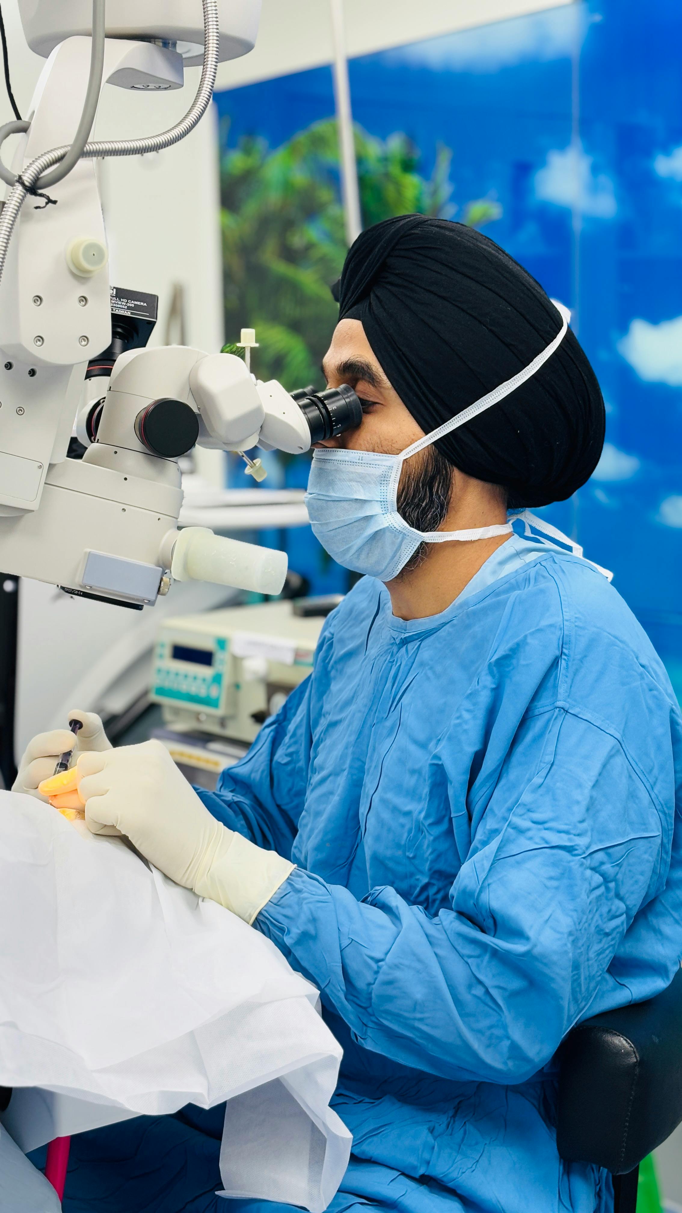
Glaucoma Surgery
Surgical procedures performed to lower intraocular pressure and protect the optic nerve in glaucoma.
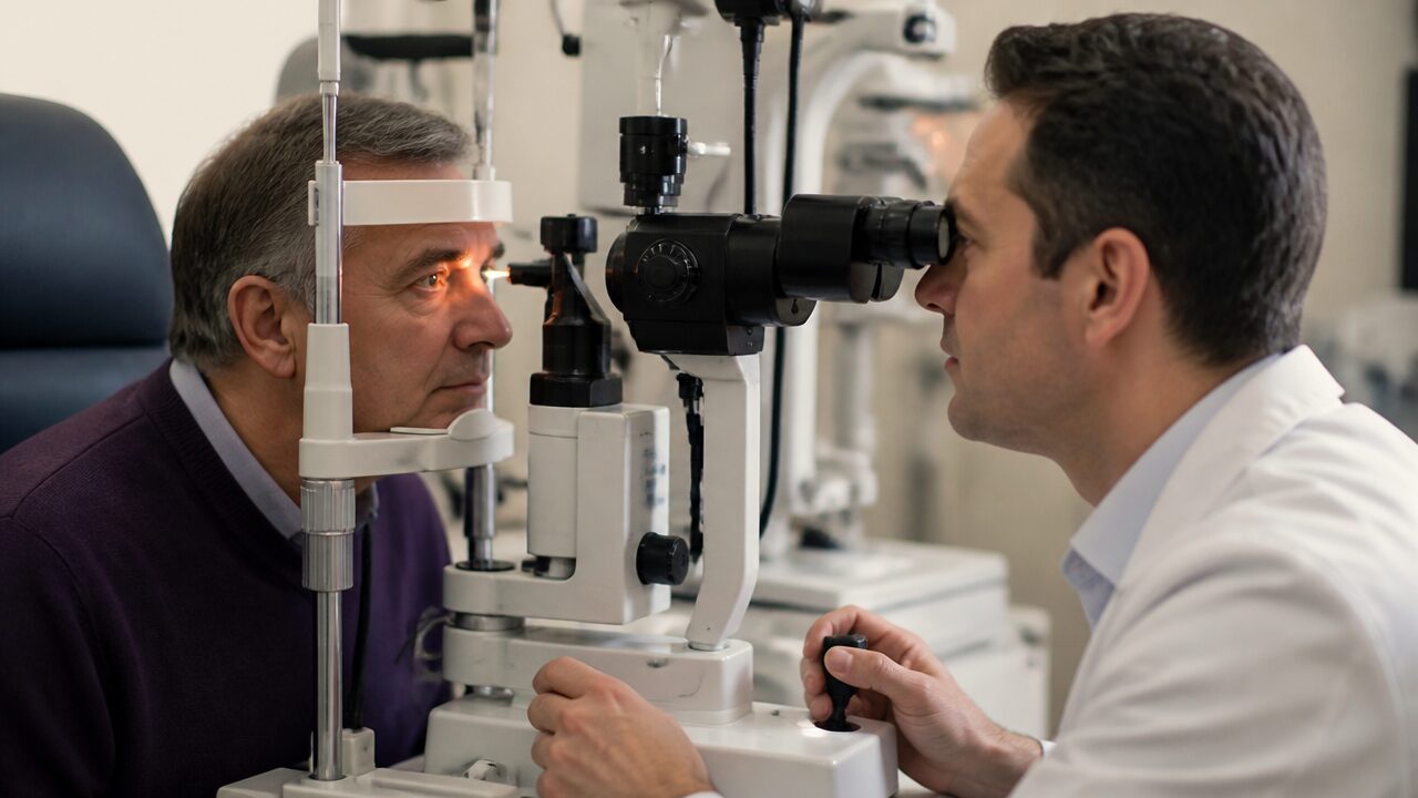
YAG Laser
A laser procedure that clears the clouded capsule behind a lens implant to restore clear vision after cataract surgery.
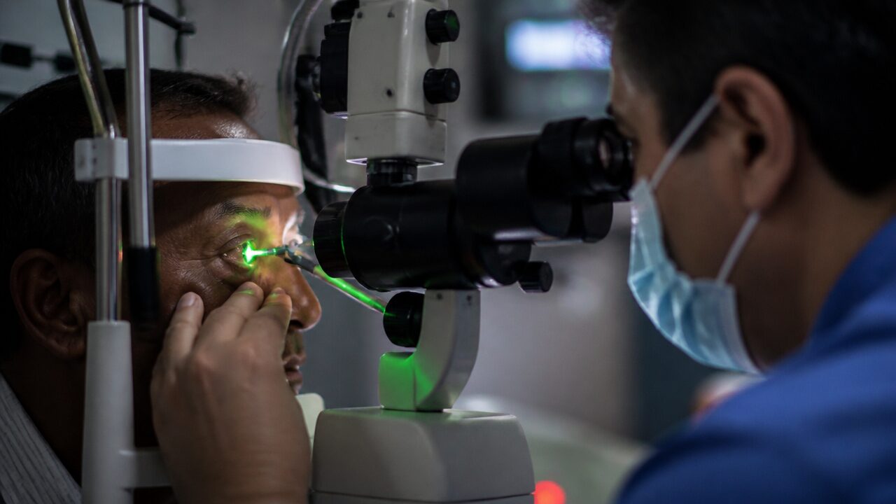
Argon Laser
A retinal laser treatment that uses controlled photocoagulation to protect vision in various retinal diseases.