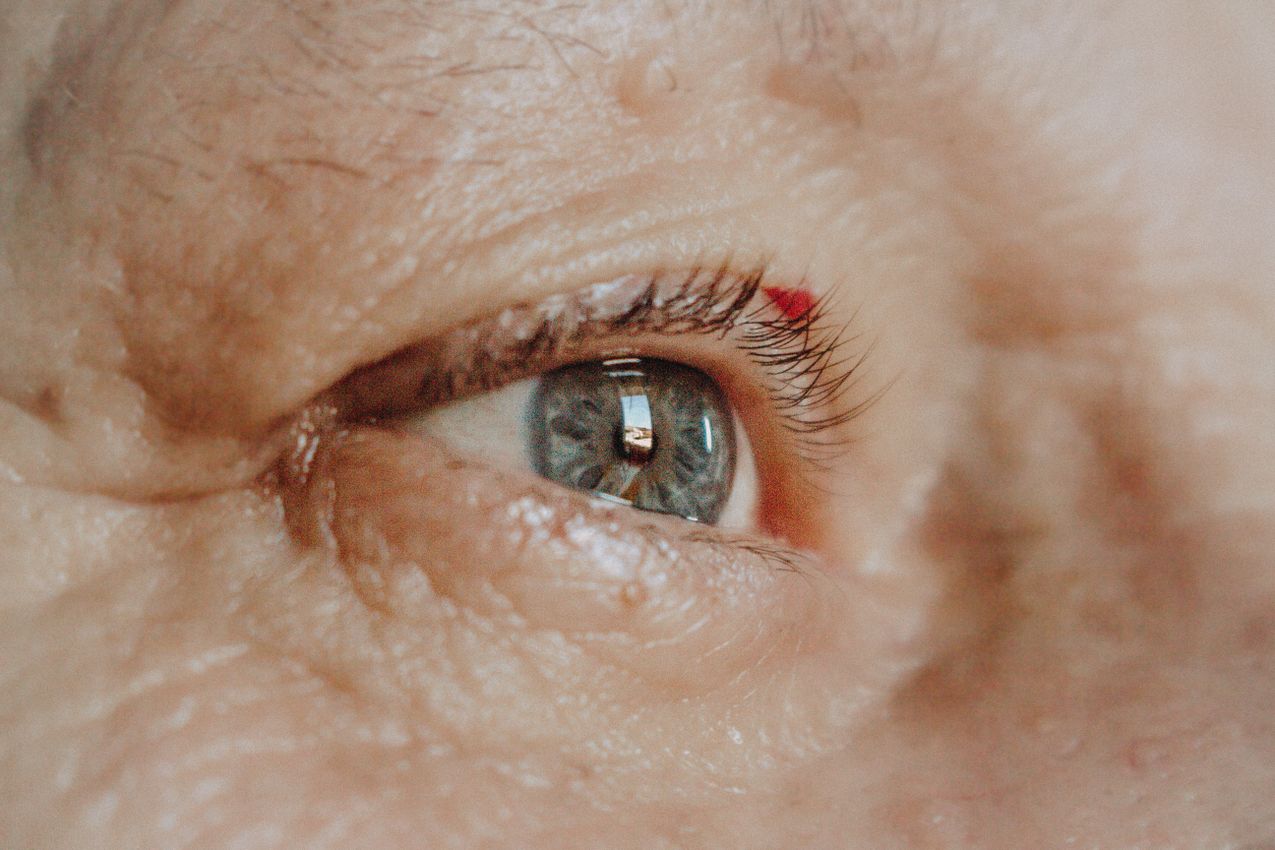
Cataracts
A cataract is the gradual clouding of the eye's normally clear lens, which reduces the quality and clarity of vision.
What Is a Cataract?
A cataract is an eye condition in which the eye's lens, normally transparent, loses its clarity and becomes cloudy. The eye's natural lens is normally clear and structured to focus light directly onto the retina. When a cataract develops, however, the lens gradually clouds over time, so less light reaches the inside of the eye and the eye's focusing power is also disturbed, leading to a serious decline in the quality of vision.
A cataract usually develops slowly and may not be noticed at first. Sometimes we see cases caused by trauma or diabetes that progress very quickly. Over time, however, a marked decrease in the sharpness of vision occurs.
Main Causes of Cataract
Ageing is the most common cause of cataract. The proteins in the eye begin to break down over the years and the lens becomes cloudy. It is seen frequently in people aged 60 and over, and the frequency rises to around 70 percent in those aged 70 and above.
Trauma can also play a role: eye injuries or eye operations can cause damage to the structure of the lens. Cataract can arise from indirect factors such as electric shock, lightning strike, radiation and heat, as well as from sharp or penetrating objects or blunt trauma.
Metabolic diseases such as diabetes can accelerate the development of cataract. Genetic factors matter too: the risk is higher in people with a family history of cataract, and congenital cataract cases also occur.
Long-term, high-dose use of steroids or cortisone can trigger cataract by speeding up the breakdown of proteins in the eye; some medications have a cataract-forming effect. Eye diseases such as uveitis and retinitis pigmentosa can also cause cataract.
Symptoms of Cataract
Cataract is usually a slowly progressing disease (though it can progress quickly with diabetes and trauma), and the symptoms generally appear over time. Our patients often do not pay much attention to it at first, because their vision is not very poor. But over time vision worsens and limits the activities of daily life.
The most common symptoms are blurred or foggy vision: as the lens clouds, images become hazy or smoky, and patients describe it as looking through a tulle curtain. There may be a change in colour perception, with colours appearing more faded or yellowed; a yellowish tone may be noticed especially in whites. Sensitivity to light is common, as bright lights, particularly sunlight or car headlights, can become uncomfortable. Difficulty seeing at night, such as trouble driving at night or seeing clearly in dark surroundings, is one of the frequently experienced problems. Frequent changes in eyeglass prescription can also occur, because the eye's refractive numbers can change quickly during cataract development, leading to the need to change glasses often.
Who Gets Cataract?
Cataract is most often seen in older people. Age-related cataract is the most common type. The reason is that, with ageing, the lens ages and loses its transparency due to causes such as increased free radicals and oxidative stress, inflammation and apoptosis. But cataract can appear even in a newborn baby just delivered (congenital cataract). The causes of newborn cataract include genetics, certain anomalies and syndromes, infections passed in the womb known as TORCH (toxoplasma, rubella, cytomegalovirus and herpes) and some metabolic diseases such as galactosemia.
Diabetes is one of the diseases in which cataract is most frequently seen. People who suffer trauma to the eye are at risk; the closer and more severe the injury is to the eye, the higher the risk. Cataract can be seen in people given cortisone (steroids) by mouth, by vein, or into the eye. Cataract is also observed more often in patients who have had uveitis or in those who are myopic. Cataract can also be seen in people who live in hot countries or work in hot occupations.
Why Does Cataract Occur?
Cataract occurs when the eye's lens loses its transparency and becomes cloudy, leading to serious deterioration in vision quality. Its causes rest on a variety of factors.
Ageing is the most common cause; as age advances, the proteins in the eye begin to break down over time and this leads to clouding of the lens, with the risk being quite high in people aged 60 and over. Genetic factors raise the risk in those with a family history, and genetic predisposition can also cause cataract to appear at an earlier age. Past physical trauma to the eye can damage the natural lens and cause clouding.
Long-term, high-dose steroid use increases the risk by causing changes in the tissues inside the eye and disturbing the structure of the eye's proteins. Diabetes and metabolic diseases can cause sugar accumulation and structural deterioration in the lens, leading to cataract at younger ages. Prolonged unprotected exposure to sunlight and UV rays can contribute to the breakdown of lens proteins, so protecting the eyes from UV is important in reducing risk.
Smoking is one of the factors that increases cataract risk, as the toxins in cigarette smoke cause oxidative stress in the eye and can damage the lens. Long-term, heavy alcohol consumption can have toxic effects on the eye's tissues and increase risk. Previous eye operations, for example retina surgery, can accelerate cataract development. And some babies can be born with cataract; these congenital cataracts usually develop in connection with genetic factors, infections in the mother (such as rubella) or metabolic diseases.
Cataract usually develops slowly over the years and emerges as a natural result of ageing, but the factors above can speed up this process or cause cataract to form at an earlier age. To protect eye health and reduce cataract risk, regular eye check-ups and the use of UV-protective glasses are important.
How Does a Person With Cataract See?
People with cataract experience a blurriness and a foggy appearance in their vision. This blurriness usually arises from the clouding over the eye's lens. As the cataract progresses, patients experience the following vision problems.
Blurred vision is the first and most common sign: patients have difficulty seeing objects clearly and vision becomes blurry, as if looking through a misted glass. As cataract progresses, colours begin to look less bright and more faded; yellowing may be noticed especially in whites, making it harder to perceive colours correctly. People with cataract are highly sensitive to bright light; dazzling light can be uncomfortable, and oncoming headlights while driving at night can be especially troublesome. Halos and glare may be seen around bright lights, which can be particularly bothersome at night, causing a ring or circular shimmer around lights. Cataract significantly affects night vision, making it hard to see in poor light. In advanced cataract, some patients may experience double vision (diplopia), especially when looking with one eye.
As an example of cataract's effect on vision: a patient reading a book may describe the letters as not appearing clear, looking blurry or running together, and may notice that colours seem dull and faded. Likewise, looking at bright sunlight or facing headlights at night becomes uncomfortable. The reason cataract leads to these kinds of vision problems is that the lens loses its transparency and prevents light from reaching the retina properly, which reduces visual sharpness and lowers vision quality.
When Should a Cataract Be Operated On?
A cataract should be operated on when it has reduced vision to the point of limiting the patient's daily-life activities and social life. This differs for each person. A pilot, a driver or a surgeon cannot tolerate reduced vision and should be operated on without delay, whereas for a homemaker or a farmer it may be possible to wait a little longer.
Before modern cataract surgery, sutured cataract surgery was performed and the cataract was allowed to harden and mature fully so that the operation would be easier. Now the situation is the opposite: we prefer not to delay the operation too much and to operate early. This is because the longer the cataract remains in the eye, the harder it becomes, the more difficult the operation is, and the complication rate increases. For this reason, please do not delay.
How Is Cataract Treated?
The method of cataract treatment is surgical. There is no medication, vitamin, drop, herb or magic formula that prevents cataract. Cataract surgery is performed by removing the clouded natural lens and placing an artificial intraocular lens (IOL) in its place. This operation is a quick and safe procedure generally carried out under eye-drop (topical) anaesthesia. With modern surgical techniques, the eye's natural lens is broken up using ultrasound by a method called phacoemulsification and removed from the eye.
After surgery, the recovery period is generally fast and patients can return to their daily activities in a short time. There may be slight discomfort, a stinging sensation or watering in the eye after the operation, but these side effects pass within a few days.
As with every surgical procedure, cataract surgery also carries some risks. Among the most common risks are eye infection, retinal detachment, or displacement of the intraocular lens.
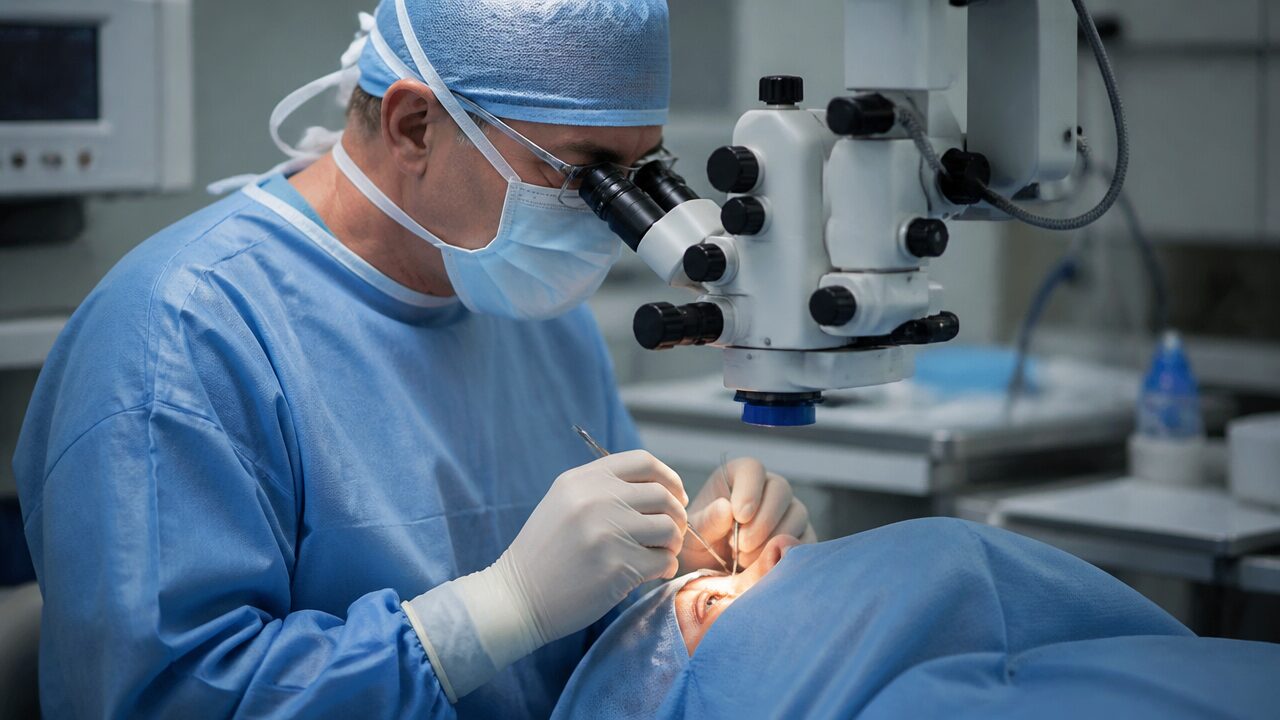
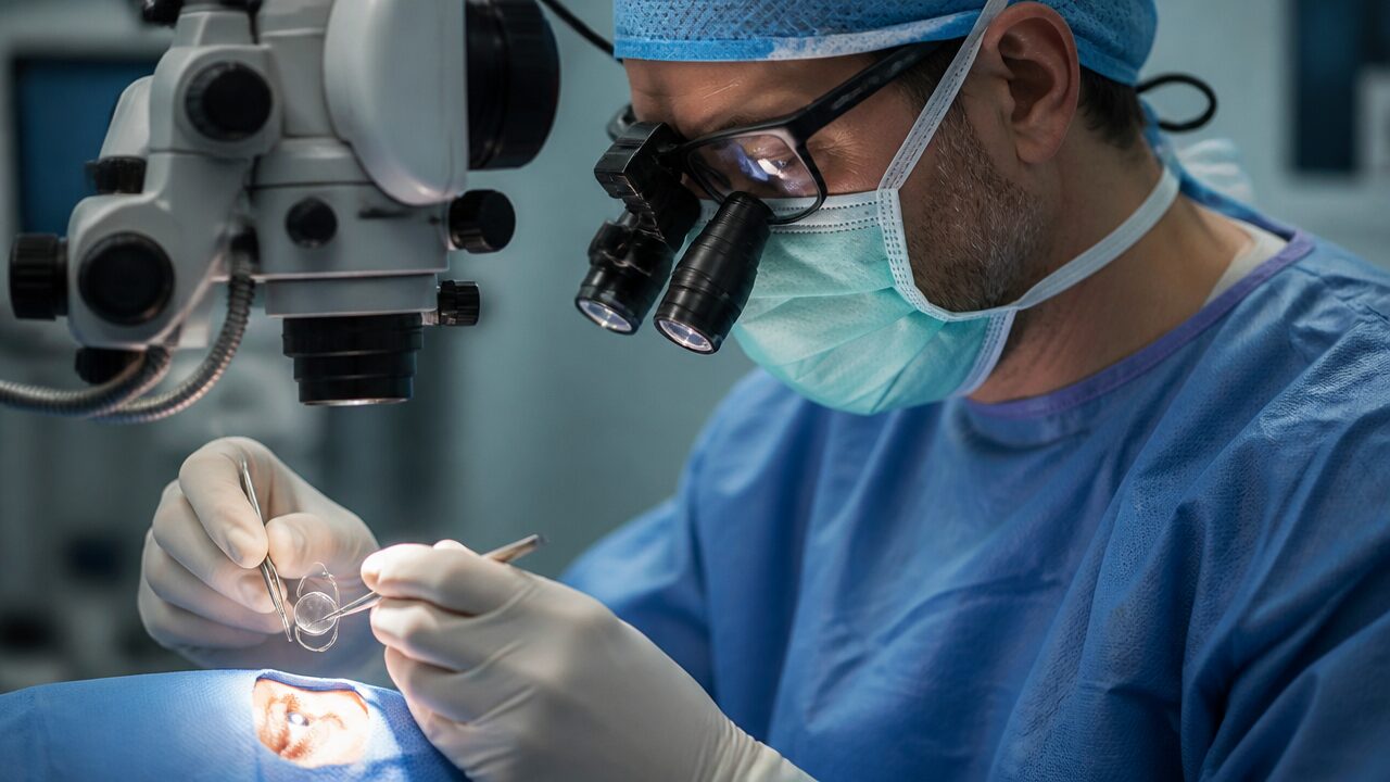
How Is Cataract Surgery Performed?
Cataract surgery is a surgical procedure in which the clouded natural lens in the eye is removed and replaced with an artificial intraocular lens (IOL). It is one of the most frequently performed operations worldwide and an effective method for restoring vision. The procedure is generally carried out in the following steps.
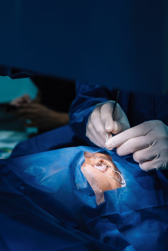
First comes preparation and anaesthesia. The operation is usually performed under eye-drop anaesthesia, we simply instil a few drops of a medication called alcaine, which keeps the patient awake while the eye is numbed, so the patient feels no pain during the procedure. No injection is given (this used to be done in the past). To keep patients comfortable during the operation, a mild sedative or sedation may be given. However, if you are a very anxious and panicky person, this operation can also be performed under general anaesthesia.
Next, a small incision is made. The eye surgeon makes a very small incision at the edge of the cornea (the clear front part of the eye), usually about 2 to 2.5 mm long, through which instruments can be placed inside the eye. When done with modern techniques, the operation generally requires no sutures. The small incision is less likely to induce astigmatism and heals quickly.
Then phacoemulsification is performed: breaking up the lens with ultrasound. This is the most widely used cataract surgery method. The cloudy lens inside the eye is broken into small pieces using ultrasound waves delivered through a probe, and the small lens fragments are suctioned out with a device called an aspirator. Performing this step as quickly, gently and carefully as possible reduces the complication rate. Some cataracts are very hard and difficult to break up; if the lens cannot be broken, the corneal incision is enlarged and extracapsular cataract extraction (ECCE) is performed.
After the natural lens is removed, a foldable artificial intraocular lens (IOL) is placed in its position. Because this lens is generally very small and flexible, it is easily inserted through the small incision made during surgery and unfolds inside the eye. The intraocular lens is generally permanent and works like a natural lens, focusing light correctly onto the retina.
Finally, the incision is closed. Because the incisions used in cataract surgery are very small, sutures are generally not needed; the corneal incision site is sealed by hydration and can close on its own, and the healing process is fast. The fluids used during the operation balance the intraocular pressure and speed healing.
After the operation, the patient is generally sent home within a few hours. On the first day there may be complaints such as blurriness, a slight stinging sensation or light sensitivity in the eye. Patients are prescribed antibiotic and anti-inflammatory eye drops to use throughout the recovery process. Healing in the eye generally takes a few weeks, but patients can usually return to their daily activities within a few days after surgery.
Femtosecond laser technology used in cataract surgery allows more precise incisions in the eye and increases the safety of the operation. With the help of this laser, it is possible to break up the cataract more precisely and to place the intraocular lens more accurately.
How Long Does Cataract Surgery Take?
Cataract surgery usually takes between 7 and 40 minutes. This time can vary depending on the surgeon's experience, the technology used and the complexity of the operation. Some eyes are very difficult to operate on (the pupil is narrow, the lens is hard, or there are accompanying problems) and this lengthens the operation time.
During the operation, anaesthesia is applied with drops, so the patient stays awake and feels no pain throughout. No injection is given into the eye in cataract surgery. Because of the pre-operative paperwork and admission, measurements, signing of consent forms, dilation of the pupil with drops, dressing in surgical clothing, blood-pressure measurement, opening of an IV line and transfer, the patient needs to arrive at the hospital one to one and a half hours beforehand. After the operation, patients are generally kept under observation for 1-2 hours and can return home the same day.
If there is cataract in both eyes, a separate operation is generally performed for each eye, with a gap of a few days or weeks between the two operations. In some very special situations the eyes may be operated on the same day.
What Is the Recovery Process Like After Cataract Surgery?
The recovery process after cataract surgery is generally fast, but full healing of the eye can take a few weeks.
In the first 24 hours, right after the operation there may be blurriness, a slight stinging sensation or itching in the eye; this is normal and decreases over time. Because of sensitivity in the eye, it may be necessary to avoid bright lights. A protective shield is recommended over the eye for the first night and perhaps a few nights, to prevent involuntary rubbing of the eye.
In the first week, the doctor prescribes antibiotic and anti-inflammatory eye drops to prevent infection risk and speed healing; these drops must be used regularly. Vision may be blurry in the first days but the eye begins to see more clearly within a few days, and in some patients full clarity may take 1-2 weeks. Patients can usually return to light daily activities within a few days, but heavy lifting, bending and strenuous exercise should be avoided.
Over the first month, vision generally improves greatly within about a week, although full healing can take 4-6 weeks, during which vision gradually becomes clearer. A few days after the operation the doctor performs a check-up to assess healing, with later check-ups generally planned at one week and one month.
There are points to pay attention to: do not touch the eye: avoid rubbing or putting pressure on it after surgery. Avoid dusty and dirty environments and protect the eye from dust, wind and water for the first few weeks to prevent infection. Contact with water and swimming are not recommended for the first few weeks; in particular, stay away from activities carrying infection risk such as swimming and pools. Avoid heavy lifting, hard exercise and bending, which can raise pressure in the eye.
In the longer term, most people can return to normal life and have greatly improved vision a few weeks after the operation. In rare cases, complications can occur during the recovery process, so it is important to consult your doctor if you notice any unexpected symptom. If blurred vision, light sensitivity or other problems continue, your doctor can carry out an assessment and recommend additional treatment. For example, some patients can develop a "secondary cataract" (posterior capsule opacification), which can be treated with a simple laser procedure. Careful care and regular doctor check-ups after cataract surgery are the key to a successful recovery.
Intraocular Lenses (IOLs) Used in Cataract Surgery
In cataract surgery, the clouded natural lens is removed and an artificial intraocular lens (IOL) is placed in its position. These lenses focus light correctly onto the retina to restore the eye's normal function. Modern intraocular lenses both improve vision quality and reduce the patient's dependence on glasses or contact lenses. However, the most suitable lens choice can differ for each patient. Here is what should be known about these lenses.
Monofocal lenses correct only a single focal point. After surgery, these lenses provide clear vision for either distance or near, and in most cases distance vision is preferred, with patients continuing to use glasses for near (such as reading). They are the most widely used lenses, carry a low complication risk, and have been used safely for many years. They are generally ideal for patients who do not mind using glasses and want clarity in distance vision.
Trifocal/multifocal lenses (smart lenses) are multi-focus lenses that can correct both near and distant distances. Because they have different focal points, they allow patients to see clearly without glasses at both near and far distances, largely eliminating the need for glasses: patients can read, use a computer or drive without glasses, though some may still need additional correction at intermediate distances (for example when using a computer). They are suitable for patients with an active lifestyle who want clear near-and-far vision without wearing glasses. However, side effects such as glare or halos around lights at night can occur in some patients.
Toric lenses are intraocular lenses designed to correct astigmatism. Patients with astigmatism experience blurred vision due to the curvature in the cornea's structure, and toric lenses correct this, treating both the cataract and the astigmatism at the same time. They reduce the need for glasses and provide clear vision at both distance and near without requiring additional treatments such as laser correction. They are the most suitable option for patients with moderate to high astigmatism; because it is very important that the lens stays in the correct position inside the eye, the surgeon's experience plays a large role.
EDoF (Extended Depth of Focus) lenses increase the depth of focus to provide clear vision over a wider range. Compared with multifocal lenses, they offer a more natural form of vision and reduce glare at night. They provide quite clear vision at distance and intermediate ranges, allowing patients to carry out daily activities without glasses, and minimise side effects such as glare. They are suitable for patients who want both near and distance vision corrected but with fewer side effects than multifocal lenses, and are especially ideal for those for whom intermediate distance (such as computer use) is important. Near vision is not as good as with trifocal lenses, and reading glasses may occasionally be used.
Aspheric lenses are designed to be closer to the eye's natural optical properties. Unlike standard lenses, they focus light more uniformly and provide clearer vision especially in low-light conditions, improving night vision and clarity in dim light by minimising light scatter in the eye. They are a good choice for patients who drive at night or have to work in dim light, and are recommended particularly for those wanting high-quality vision.
Points to Consider When Choosing an Intraocular Lens for Cataract Surgery
The lens choice should be made according to the patient's lifestyle. For patients with an active lifestyle who do not want to wear glasses, multifocal or EDoF lenses may be more suitable. However, for patients who frequently drive at night, aspheric lenses may be preferred over multifocal lenses, which can make them sensitive to glare.
For patients with astigmatism, toric lenses are the most suitable option, as these lenses treat both the cataract and the astigmatism at the same time. Each lens type can present different side effects: the technology is already good but still not perfect: multifocal lenses can cause glare at night, while EDoF lenses can offer a more natural visual experience. It is important for patients to discuss this matter in detail with their doctor.
What Are the Risks and Complications of Cataract Surgery?
Cataract surgery is today one of the most frequently performed surgical procedures. However, as with every surgical intervention, cataract surgery can also have some complications. Volumes upon volumes of books have been written about these complications, and some of them can even lead to loss of the eye. Although these risks are generally low, it is important for patients to be informed about these complications before the operation.
Posterior capsule opacification (secondary cataract): during cataract surgery, while the natural lens is being removed, the front capsule of the lens is peeled off but the back capsule is left in place. The reason is to keep the lens stable and prevent it from falling backward; this capsule is used to support the artificial intraocular lens (IOL). However, months or years after the operation, cell accumulation can occur in this capsule, causing vision to become blurry again. This occurs in about 30-40 percent of all operated patients and is more frequent in those who had cataract surgery as children or at a young age. Although patients often think of it as "the cataract coming back," it is not truly a cataract. It can be treated with a laser procedure called YAG laser capsulotomy that takes about 3-5 minutes.
Infection (endophthalmitis): there is a risk of infection developing in the eye after surgery. This condition is called endophthalmitis and shows itself with severe inflammation inside the eye. It is a rare but serious complication, appearing with symptoms such as eye pain, redness and severe blurred vision. If not treated early, the patient can lose the eye. If the infection is noticed early, treatment with strong antibiotics and antifungal medications is possible, but in advanced cases it can lead to vision loss.
Retinal detachment: after cataract surgery, the risk of retinal detachment can increase, especially in myopic patients. The retina is the layer at the back of the eye that provides vision. Changes in intraocular pressure during the operation can cause the retina to detach. Patients experiencing retinal detachment notice flashes of light, sudden vision loss or a feeling like a curtain coming down over the eye. It is a situation requiring urgent surgical intervention and can lead to permanent vision loss if left untreated.
Chronic raised eye pressure (ocular hypertension): in some patients, intraocular pressure can rise after the operation due to blockages in the eye's drainage system or inflammation. Pressure-lowering drops are used, but if the pressure cannot be brought under control there is a risk of glaucoma developing.
Cystoid macular oedema: fluid accumulation in the eye after surgery can cause swelling, especially in the macular region; this is called cystoid macular oedema (CME) and can blur central vision. Patients usually present with complaints of blurred or distorted vision. It is treated with steroid or non-steroidal anti-inflammatory eye drops.
Intraocular lens dislocation: displacement of the artificial intraocular lens (IOL) placed during the operation is possible, though rare. A lens in the wrong position can affect vision quality and cause blurred or double vision. Small position changes can be monitored, but if the lens fully dislocates, surgical intervention may be required.
Corneal oedema (swelling): swelling can be seen in the cornea after surgery, occurring especially when the cornea is traumatised during the operation. Loss of corneal cells can cause the cornea to lose its clarity and lead to blurred vision. In mild cases the condition can resolve on its own, but in more serious cases intensive treatment or a corneal transplant may be needed.
Increase in astigmatism: after surgery, some patients can develop astigmatism or have existing astigmatism increase, depending especially on how the incision area of the eye heals. It can be treated with glasses, contact lenses or laser correction.
Halos and glare around lights: after surgery some patients may notice halos or glare around lights, which can create difficulty especially in night vision. These side effects usually decrease over time, but can be permanent for some patients.
Certain conditions increase risk factors. Diabetic patients can be more prone to complications after cataract surgery. Patients with high-degree myopia can have an increased risk of retinal detachment. Patients who have had previous eye surgery have a higher complication risk.
In conclusion, cataract surgery is generally a safe procedure that greatly improves vision quality. However, being informed about these complications calls for careful follow-up and prompt intervention after the operation. Regular follow-ups with an eye-health specialist are important for the early diagnosis and treatment of possible complications.
Can Cataract Be Prevented?
The formation of cataract cannot be completely prevented, but it is possible to reduce the risk factors. Protection from UV rays, a balanced diet, quitting smoking and having regular eye check-ups can slow the development of cataract. In addition, keeping metabolic disorders such as diabetes under control can lower cataract risk, and the use of cortisone should be limited.
For years, patients of mine who saw well have a hard time accepting reduced vision when they get a cataract. I tell them this: just as your skin wrinkles and your hair turns white, your lens also ages and loses its transparency. But then, once the talk of age comes up, they get cross with me. Still, what can we do: the words that best explain cataract are 'the ageing of the lens.' Should we not tell the truth too?
Types of Cataract
Cataract is an eye disease that arises from clouding of the eye's lens, but not every cataract case is the same. There are different types, classified according to which region of the lens they form in and how the cataract develops.
Nuclear sclerotic cataract forms in the centre (nucleus) of the eye's lens and develops as part of the ageing process. Over time the lens hardens and takes on a yellowish colour. At first there may be a temporary improvement in near vision (for example, people may be able to read without reading glasses), but as it progresses, problems such as blurred vision and colours appearing less bright arise. It is generally common in people aged 60 and over.
Cortical cataract is characterised by white or grey wedge-shaped opacities that begin at the outer edge of the lens (the cortex) and extend toward its centre, preventing light from entering the eye properly. People with cortical cataract generally experience glare, dazzling and difficulty especially when driving at night. This type is seen more often in people with diabetes.
Posterior subcapsular cataract forms at the back of the lens (the back capsule) and prevents light from reaching the retina properly. This type can progress rapidly and can produce symptoms in a shorter time than other types. Extreme sensitivity to light, a sudden deterioration in near vision and halos around bright lights are common. It can develop in diabetic patients, people who have used steroids long-term, in trauma and at young ages.
Congenital cataract: some babies can be born with cataract or develop it in the first years of life. It can occur due to genetic factors, infections in the womb (toxoplasma, rubella, cytomegalovirus, herpes) or metabolic diseases (galactosemia). It can be detected during a child's eye examination and can lead to vision loss if early intervention is not made. It arises from genetic predisposition or infections the mother had during pregnancy (for example rubella).
Traumatic cataract arises from disruption of the lens structure after trauma or injury to the eye. It can develop from a sudden blow, chemical burns or exposure to radiation, and can show sudden vision loss, clouding or light sensitivity. It can be seen in people who have suffered eye injuries.
Secondary cataract develops after cataract surgery in some patients as a result of cell accumulation in the back capsule of the lens. It is called "secondary cataract," but it is not actually a true cataract, and is generally treated with a procedure called YAG laser capsulotomy. The complaint of blurred vision can occur again after the operation.
Preparations Before Cataract Surgery
Cataract surgery is generally a short and safe procedure, but preparations before the operation can speed recovery. There are some important steps for patients to pay attention to before surgery.
A detailed eye examination comes first. In the detailed examination before the operation, the artificial intraocular lens (IOL) most suitable for your eye's structure and visual needs is selected: this may be a monofocal, bifocal, EDoF, monofocal plus, multifocal, or astigmatism-correcting toric lens. Your doctor will make the best lens choice taking into account your needs in daily life. Eye measurements are also taken: intraocular pressure, eye structure and other important parameters are measured to determine the technique to be used during surgery.
Regarding medications, the medicines you use before the operation are reviewed by your doctor. Blood-thinning medications are not stopped before the operation. Antibiotic eye drops may be prescribed to reduce infection risk during and after surgery.
General health condition is also assessed. Your doctor may request blood tests to learn about your general health; blood sugar is checked for diabetic patients, and tests for hepatitis B, hepatitis C and HIV (called ELISA) should be performed for every patient. Factors such as high blood pressure can also affect the surgical process, so regular check-ups are important. If you are diabetic, make sure your blood sugar level is under control, as high blood sugar can slow healing; generally a blood sugar below 200 is desired before the operation.
On the day of surgery there are preparations to make. Fasting is normally not needed; in fact, a light breakfast before the operation is recommended. If the operation is to be done under general anaesthesia, you generally need to eat and drink nothing for 6-8 hours beforehand, to ensure an empty stomach during anaesthesia. You must have your eyes completely free of any make-up or cream on the day of surgery to minimise infection risk. Because there will be blurriness and slight discomfort in the eye after the operation, you may need a companion to go home and rest; driving is not allowed after surgery.
Psychological preparation matters too. Before the operation some patients may feel tension and anxiety; in that case your doctor can give a sedative medication. Because cataract surgery is performed under drop anaesthesia, you will be completely awake but will feel no pain, and knowing that the operation is quick can help you relax. Proper preparation makes recovery after cataract surgery smoother; you can create a preparation plan specific to you by talking these matters over with your doctor before the operation.
Are Blood Thinners and Other Medications Stopped Before Cataract Surgery?
Unless your doctor says otherwise, no medication needs to be stopped. Cataract surgery is generally done with drop anaesthesia and no injection is given into the eye, so blood thinners are not stopped. However, in some situations local anaesthesia may be required, or when retina surgery (vitrectomy) is performed together with cataract surgery, it may be necessary to stop blood thinners. Be sure to ask your eye doctor about this.
Lifestyle After Cataract Surgery
Although cataract surgery is a successful surgical intervention, it may be necessary to take some precautions during recovery and make certain changes in lifestyle. The points to pay attention to after surgery are important for protecting eye health and speeding healing.
Use of eye drops is essential: the antibiotic and anti-inflammatory eye drops given by the doctor should be used regularly to reduce infection risk and keep inflammation under control after surgery; drop use generally lasts a few weeks. The drop-use period must be followed and the treatment not interrupted for as long as the doctor specifies; also take care not to instil drops into your eyes without washing your hands.
Protecting the eye is important. The doctor may recommend wearing a protective eye patch or shield for a few days after surgery to protect the eye from blows or rubbing, which is especially important while sleeping to prevent involuntary rubbing of the eye. Sensitivity to sunlight and bright lights can increase in the first few weeks, and wearing quality UV-protective sunglasses when outdoors during this period helps protect your eyes.
Physical activity and rest also matter. During the first week after surgery, heavy lifting and bending should be avoided, as such movements can raise intraocular pressure and adversely affect recovery. Heavy exercise such as swimming and running should be avoided for the first few weeks; activities such as light walks can be done until the eye heals, but it is important to get your doctor's approval. Pay special attention to resting and not tiring your eyes during the first week, and it can be helpful to keep activities such as reading and watching television limited.
Hygiene and eye contact require care. Slight itching or discomfort in the eye after surgery is normal, but rubbing your eyes can damage the recovery process, increase infection risk and slow healing. Avoid direct contact of your eyes with water for the first few weeks; in particular, avoid activities such as swimming and protect the eyes while showering.
Regarding glasses, your eye prescription may change after surgery, so a new glasses prescription may be needed after a while; however, you should wait for your eyes to heal fully before new glasses, which generally takes a few weeks. Some patients may use temporary glasses for a short time after surgery, and your doctor will guide you on this.
Check-ups during recovery are part of the process: after surgery your doctor will carry out check-up examinations at certain intervals, usually the first within a few days, followed by regular follow-ups at one week and one month, during which the eye's healing is monitored and possible complications can be detected early.
There may also be visual differences during recovery. In the first few days after surgery there may be blurriness or slight distortion in vision; this is usually temporary and improves as the eye heals, and it can take a few weeks for the new lens to fully adjust with the eye. After cataract surgery, patients generally notice that colours appear brighter and clearer, which is normal because the cloudiness of the eye's natural lens has been removed. Following the doctor's advice and paying attention to the recovery process are key to achieving a successful result.
What Can I Do to Slow Down and Prevent Cataract?
Cataract is an age-related eye disease and is not a fully preventable condition. However, there are some precautions that can be taken to slow its development and protect eye health. These measures, together with lifestyle changes and regular eye check-ups, can minimise cataract risk.
Protection from UV rays is important: exposure to the sun's harmful UV rays can increase the risk of cataract, so wearing quality UV-protective sunglasses when going out in the sun is one of the most important ways to protect your eyes: especially if you spend long periods outdoors, you should apply this regularly. Wearing a wide-brimmed hat also provides an additional advantage in protecting your eyes from the sun.
Healthy nutrition helps: cataract development is associated with oxidative stress in the eye, and an antioxidant-rich diet can reduce this stress. It is especially important to consume foods containing vitamin C, vitamin E, beta-carotene and lutein, which protect eye health and minimise the harm from free radicals. Consuming green leafy vegetables such as spinach and kale, and fish rich in omega-3 fatty acids such as salmon and tuna, is beneficial for eye health.
Quitting smoking and alcohol use is beneficial. Smoking causes oxidative stress in the eye and increases cataract risk, and it is one of the most important lifestyle factors that accelerate cataract development; quitting smoking reduces not only cataract risk but also the risk of other eye diseases. Excessive alcohol consumption can also harm eye health and increase cataract risk, so limiting alcohol intake contributes to eye health.
Regular eye check-ups are important: because cataract progresses slowly, early diagnosis allows intervention before vision loss occurs. It is especially important for people over 40 to have regular eye examinations, which play a key role in the early diagnosis of cataract. During eye examinations, besides cataract, other age-related eye diseases such as glaucoma and macular degeneration can also be detected.
Diabetes control matters: the risk of cataract is higher in diabetic patients, and keeping blood sugar levels under control helps prevent the eye diseases caused by diabetes, so it is very important for diabetic patients not to neglect regular eye check-ups.
A healthy lifestyle helps too. Getting enough sleep is important for eye health, allowing your eyes to rest and renew themselves. Regular exercise can help protect eye health, as physical activity increases blood circulation and ensures enough oxygen and nutrients reach your eyes. Limiting steroid use is also important: long-term steroid use can cause cataract development, so using medications under a doctor's supervision is important; if you use steroids, it is helpful to consult your doctor about possible side effects and to have more frequent eye check-ups.
Although these measures cannot completely stop cataract development, they can reduce and delay the risk of cataract formation. Regular eye check-ups and healthy lifestyle habits have a protective effect against cataract and other eye diseases.
Treatment Options for Better Vision After Cataract Surgery
Cataract treatment generally ends with a surgical intervention. However, to obtain better vision results after the surgical procedure, some options can be offered to patients. These can minimise patients' need for glasses or lenses and improve vision quality.
Trifocal (multifocal) lenses (smart/premium lenses) correct both near and distance vision problems. While standard monofocal lenses can correct only a single distance, multifocal lenses have different focal points and so provide clear vision at near, intermediate and far distances. Their biggest advantage is that they largely reduce the patient's need for glasses after surgery, improving quality of daily life: activities such as reading, using a phone or driving can be done without glasses. They are ideal especially for patients who want a glasses-free life, but may not be suitable for patients with other eye problems or very high astigmatism; occupation, age and the anatomical features of the eye are very important in choosing this lens.
Toric lenses are specially designed to correct astigmatism. For patients with astigmatism, normal lenses may be inadequate, and toric lenses then come into play. Placed during the operation, they correct astigmatism to a large extent and reduce the need for glasses, so patients can have clearer distance and near vision after surgery. They are one of the most suitable options for patients with astigmatism, but should still be applied in line with the doctor's recommendation.
Laser-assisted cataract surgery is a modern technology; while traditional cataract surgery is done manually, in laser-assisted surgery the incisions and the breaking up of the lens are done with laser technology, providing a more precise and controlled operation. It can reduce complication risk and may speed the recovery process, and the laser can also be used to correct astigmatism. It is ideal for patients who want a more precise operation; however, this method can be more expensive than traditional surgery, and for this reason it has not become very widespread.
Monovision is a method in which one eye is set for distance vision and the other for near vision, and can be used as an alternative to multifocal lenses. It can be more affordable than multifocal lenses and allows patients to live without glasses at both far and near distances, although the brain must adapt to each eye focusing at different distances. It is a good alternative for patients who are not suitable for multifocal lenses but want glasses independence, and still gives very good results in some selected patients.
YAG laser capsulotomy: after cataract surgery, opacification can develop in the back capsule of the lens (secondary cataract) in some patients, and this can be treated with a simple laser procedure called YAG laser capsulotomy. The YAG laser is a procedure that can be done in a very short time and painlessly, and is an ideal solution for patients with blurred vision again after surgery. It can be applied to all patients who develop posterior capsule opacification after cataract surgery.
Frequently Asked Questions
Is pain felt during cataract surgery? No, you do not feel pain during cataract surgery. The operation is done with topical or local anaesthesia, that is, the eye is numbed with eye drops, so the patient stays awake but feels no pain or discomfort. Only a slight pressure may be felt around the eye, but this is generally not uncomfortable.
Will I need to use glasses after cataract surgery? Because the monofocal lenses used in standard cataract surgery correct only distance or only near vision, you may need reading glasses after surgery. However, if you choose trifocal, multifocal, EDoF or monofocal plus lenses, they can correct both near and distance vision and largely eliminate the need for glasses.
When can I return to work after cataract surgery? Resting for the first few days after the operation is recommended. Once your eye has healed, you can generally return to your daily activities and work within a day or two. However, those who work in heavy jobs or in dusty and crowded environments may need to be careful for a longer time.
Is cataract surgery risky? Cataract surgery is today one of the safest surgical procedures. However, as with every surgery, rare complications such as a low risk of infection, retinal detachment or a rise in intraocular pressure can occur. For this reason, regular check-ups after surgery and early intervention at any problem are important.
How long does it take for my vision to improve after cataract surgery? Vision quality increases quickly after the operation. Blurred vision in the first few days is normal, but the eyes generally begin to heal within a week and vision becomes clearer. The full recovery process can take 4-6 weeks.
Can cataract surgery be repeated? Cataract surgery is generally permanent and does not need to be repeated. However, in some patients a "secondary cataract" can develop after the operation, and in that case it is possible to resolve the problem with a simple laser procedure called YAG laser capsulotomy.
Can the lens shift after cataract surgery? Although very rare, displacement of the artificial lens placed in the eye is possible. In that case there can be deterioration in vision and surgical intervention may be required. Such situations generally depend on the surgeon's experience and on following the points to pay attention to after surgery.
When can I bathe or swim after cataract surgery? After the operation, the eyes need to be protected from water in the first weeks while they heal. Activities such as swimming are generally not recommended for 3-4 weeks, but the duration is set according to your doctor's advice.
Can I drive after cataract surgery? Because there may be blurred vision right after the operation, driving can be dangerous. Driving should be avoided until the eyes heal and vision becomes clear (generally a few days to a week).
Does cataract develop again after cataract surgery? Cataract does not form again after cataract surgery. However, in some patients a condition referred to as "secondary cataract" can develop after the operation; this is not actually a true cataract. During the operation a large part of the front capsule of the lens is removed but the back capsule is left in place, and opacity can develop on the back capsule where the intraocular lens is placed. Posterior capsule opacity can occur at varying rates and times depending on the patient's age, any pre-existing eye disease, the presence of uveitis, whether the cataract was trauma-related, and the lens placed. In this case the patient reports that their vision has decreased months or years after the operation, and this opacity is cleared with YAG laser capsulotomy. This procedure is not surgery, is painless, and is done on an outpatient basis within 1-5 minutes; it is done once. We generally explain it to our patients as something like 'calcification' or 'soiling.' In reality, it occurs when lens epithelial cells that are stuck to the back capsule and invisible to the eye multiply over time, spread over the back capsule and cover the visual axis. The 'calcification' does not recur, and the patient thus regains their former vision. Although it is a short procedure, because it removes the wall between the front and back chambers of the eye, retinal tears and detachment, intraocular lens displacement or a rise in intraocular pressure can rarely be seen.
This page is for general information and does not replace a personal examination. The right approach is decided together after an eye examination.
Cataract Surgery
A short, common procedure in which the clouded natural lens is removed and replaced with an artificial intraocular lens to restore vision.
Smart Lens Surgery
A procedure that replaces the eye's natural lens with an artificial smart lens to correct presbyopia and cataract and reduce dependence on glasses.
Scleral-Fixated Lens Surgery
A procedure that secures an intraocular lens to the sclera when there is no capsular support to hold it in place.
YAG Laser
A laser procedure that clears the clouded capsule behind a lens implant to restore clear vision after cataract surgery.