Scleral-Fixated Lens Surgery
A procedure that secures an intraocular lens to the sclera when there is no capsular support to hold it in place.
Scleral-fixated lens surgery is a surgical procedure in which an intraocular lens is placed and fixed to the sclera (the white part of the eye). It is usually used in situations where the natural intraocular lens has been removed (after cataract surgery or trauma) but there is not enough capsular support to hold the lens in place. This surgery aims to provide clear vision by keeping the lens fixed in the eye. This is not something done routinely, because normally artificial intraocular lenses are placed in the position of the natural lens removed from the eye, within the structure called the 'bag', between the posterior and anterior capsule. But if for any reason this support in the eye has been lost, then the lenses are either sutured to the sclera or the haptics (legs) of the lens are buried into the sclera. A scleral-fixated lens helps the lens to be firmly fixed inside the eye in such situations.
In Which Situations Is Scleral-Fixated Lens Surgery Needed?
Scleral-fixated lens surgery is preferred in situations where there is no capsular support to fix the intraocular lens. This surgery is usually performed in the following situations. Loss of capsular support: if the capsular bag is damaged or inadequate during cataract surgery, scleral fixation is needed to place and fix the lens. Trauma: if the lens support structures are damaged as a result of eye injuries, scleral-fixated lens surgery may be needed. Lens dislocation: in cases where the natural lens or a previously placed intraocular lens slips out of position (dislocation), scleral fixation may be needed. Lens implantation: this technique can be used for lens implantation in patients who have no lens capsule from birth.
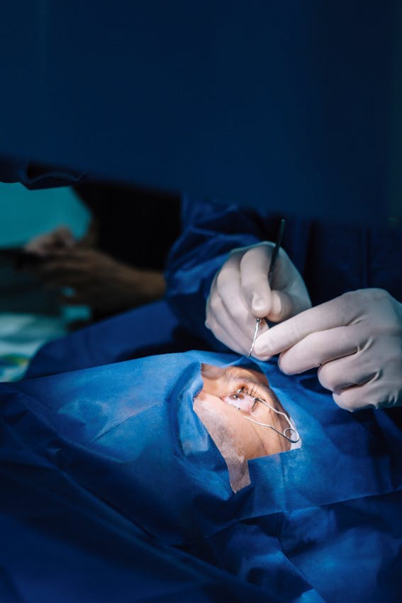
How Is Scleral-Fixated Lens Surgery Performed?
Scleral-fixated lens surgery involves fixing the intraocular lens to the sclera (the white part of the eye) with sutures, or burying its haptics. The surgery is performed by an experienced eye surgeon and is usually done under local anaesthesia. The procedure is carried out in the following steps.
Stages of the Surgery
Anaesthesia: before the surgery the area around the eye is numbed with local anaesthesia, so the patient does not feel pain or discomfort during the procedure. In some cases general anaesthesia may also be used.
Lens placement: the eye surgeon places an artificial intraocular lens (IOL) inside the eye. This artificial lens is usually a lens-shaped implant and provides the patient's clear vision.
Scleral fixation: to fix the lens to the sclera, the surgeon makes small holes in the sclera. The artificial lens is fixed with sutures placed in these holes. Thanks to the sutures, the lens is firmly settled inside the eye and does not move.
Final check and closure: after the surgeon is sure that the lens is in the correct position, the incisions in the eye are carefully closed. A sterile dressing is applied before the eye enters the healing process.
Recovery After Surgery
The recovery time after scleral-fixated lens surgery can vary depending on the patient's general health and the extent of the surgery. However, most patients can return to their daily lives within a few weeks.
The First Days
Mild discomfort in the eye: after surgery there may be mild pain, burning and a feeling of discomfort in the eye. This usually decreases within a few days. Vision changes: in the first days after surgery vision may be blurry, but this improves along with the healing process. Swelling and redness: there may be mild swelling and redness around the eye, which disappears within a few days.
Care and Use of Eye Drops
Eye drops: after surgery your doctor may prescribe eye drops and ointment. These drops are used to prevent infection and speed up healing. Protecting the eye: to protect the eye from infection or trauma, it may be necessary to wear an eye patch or glasses for a few days.
Check-ups
After surgery your doctor will plan several follow-up appointments. At these check-ups the position of the lens inside the eye and the healing process are monitored.
Advantages of Scleral-Fixated Lens Surgery
Fixation of the lens: fixing the intraocular lens to the sclera prevents the lens from slipping or shifting inside the eye. Providing clear vision: with the fixation of the lens, the surgery aims to provide the patient's clear vision. Long-term solution: scleral fixation offers a lasting solution by keeping the intraocular lens securely fixed for a long time.
Risks of the Surgery
Like any surgical intervention, scleral-fixated lens surgery also carries some risks. However, these risks are usually low. Infection: there is a risk of infection in the eye after surgery, but the antibiotics recommended by the doctor reduce this risk. Bleeding: there may be minimal bleeding around the eye. Increased intraocular pressure: rarely, intraocular pressure may rise after surgery. This should be monitored carefully and treated if necessary. Visual disturbance: in very rare cases, a visual disturbance may occur after surgery, but this usually improves over time.
Don't Delay Treatment
Scleral-fixated lens surgery is an effective treatment method for restoring vision in patients who have no intraocular lens support. If you are having a problem with your intraocular lens, it is important to consult an eye doctor about this surgery to protect your eye health and to provide clear vision.
This page is for general information and does not replace a personal examination. The right approach is decided together after an eye examination.