Tear Duct Obstruction Surgery
A surgery that opens a blocked tear drainage channel so tears can flow normally from the eye into the nose again.
What is tear duct obstruction?
Tear duct obstruction (nasolacrimal duct obstruction), or chronic dacryocystitis, is the condition in which the tears produced in the eyes cannot pass through the tear duct that drains toward the nose. Normally, tears are produced continuously to keep your eyes moist and healthy and flow through a channel from two small openings on the inner part of the eye toward the nose, then into the nose and finally into the throat. However, when this channel becomes blocked, tears cannot flow normally and problems such as watering, infection, and inflammation arise in the eyes. Tear duct obstruction can be congenital, or in adults it can develop due to causes such as aging, injuries, infections, or tumors. In most cases surgical intervention is required, and this operation generally gives successful results.
What is tear duct obstruction surgery?
Tear duct obstruction surgery is known as dacryocystorhinostomy (DCR), and it opens the blockage in the tear duct to restore the normal drainage of tears. This operation is performed to create a new pathway that allows tears to flow into the nose. This new passage in the tear duct eliminates problems such as watering, infection, and discomfort caused by the blockage.
Symptoms of tear duct obstruction
The symptoms of tear duct obstruction usually include constant tearing (epiphora), watering and irritation in the eyes, redness around the eye, frequent infections in the eye, crusting and discharge in the eye, and a runny nose (especially with an infected tear duct). These symptoms are usually bothersome and can negatively affect daily life. If you are experiencing one or more of these symptoms, you may have a tear duct obstruction.
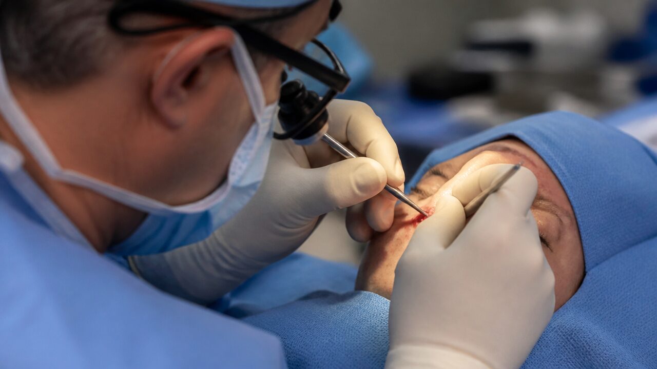
How is tear duct obstruction surgery performed?
A permanent solution for tear duct obstruction usually requires surgical intervention. This surgery, called dacryocystorhinostomy (DCR), is performed to open the tear ducts and allow tears to drain properly into the nasal cavity.
Stages of the operation
Anesthesia: the operation is usually performed under local anesthesia, but in some cases general anesthesia may also be preferred. This depends on the patient's condition and the surgeon's preference.
Surgical methods
External dacryocystorhinostomy: in this traditional method, a small incision is made in the skin between the nose and the eye. Through this incision the tear duct is opened and tears are allowed to flow easily into the nasal cavity. The incision is then closed with sutures, and once the wound heals the scar usually becomes indistinct. Endoscopic (laser) dacryocystorhinostomy: this modern method is carried out from inside the nose with the help of an endoscope or laser, without making an incision in the skin. The blockage in the tear duct is opened from the inside of the nose and the treatment is completed without a scar. Use of a temporary tube or stent: after opening the blockage, the surgeon may temporarily place a tube or stent in the tear duct. This stent keeps the tear duct open and is removed after a few weeks. Completion of the operation: after the operation is completed, patients can usually return home the same day. The recovery time varies depending on the surgical method and the patient's general health.
The recovery process after surgery
The recovery process after tear duct obstruction surgery is usually completed within a few weeks. The following points should be noted after the operation.
The first days
Swelling and bruising: there may be mild swelling and bruising around the eye after the operation; this usually subsides within a few days. Mild discomfort in the eye: mild pain and discomfort may be felt after the operation, and your doctor may prescribe pain relievers to ease this discomfort. Vision changes: there may be blurred vision and watering in the eye, but these symptoms are temporary and disappear during the healing process.
Care and medication use
Eye drops and ointment: antibiotic eye drops or ointment should be used after the operation to prevent infection and speed up healing. These medications should be used regularly as recommended by the doctor. Eye hygiene: the area around the eye should be kept clean and the hygiene rules recommended by the doctor should be followed.
Follow-up examinations
After the operation your doctor monitors the condition of the tear duct with regular check-ups. If a tube or stent was placed during the operation, these materials are removed during the healing process.
The benefits of the operation
Protection of eye health: tear duct obstruction surgery allows tears to drain properly into the nasal cavity and eliminates the risk of infection caused by tears accumulating in the eye. Relief in the eye: after the operation, the constant tearing (epiphora) is eliminated and patients are relieved of the bothersome sensation of watering in the eye. Prevention of infections: the infections that frequently arise due to the blockage are eliminated after the operation.
The risks of the operation
Tear duct obstruction surgery is generally a safe procedure, but as with any surgical procedure there are some risks. Infection: the risk of infection after surgery is rare, but it can be prevented with antibiotic treatment. Bleeding or swelling: temporary bleeding, bruising, and swelling may be seen around the eye, but this usually resolves in a short time. Recurrence of the blockage: in rare cases the tear duct may become blocked again, in which case a second surgery may be required. Dryness in the eye: rarely, opening the tear duct too much can lead to dryness of the eye.
Do not postpone tear duct obstruction surgery
If left untreated, tear duct obstruction can lead to frequently recurring infections, eye irritation, and even vision loss. If you are experiencing symptoms such as constant watering, infection, or redness around the eye, it is important to consult an ophthalmologist. Early intervention quickly resolves the problem and protects your eye health.
This page is for general information and does not replace a personal examination. The right approach is decided together after an eye examination.