Pterygium Surgery
Surgical removal of a pterygium, a fleshy growth that extends from the white of the eye toward the cornea.
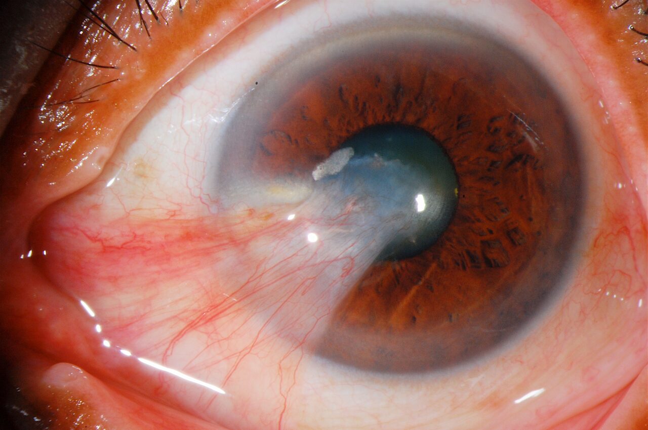
Pterygium surgery is the procedure of surgically removing a pterygium, known as a "flesh growth" or "bird's wing," which begins on the white part of the eye (conjunctiva) and extends toward the cornea. A pterygium usually forms with environmental factors such as excessive sun exposure, wind, and dust, or with genetic factors, and if left untreated it can cover the field of vision and cause vision loss. Pterygium surgery is performed for both aesthetic and functional reasons.
What Is a Pterygium?
A pterygium is a condition characterized by abnormal growth of the conjunctival layer of the eye. It begins on the white part of the eye (sclera) and advances toward the cornea (the transparent part of the eye). It is usually seen in people exposed to UV rays for a long time. When a pterygium progresses, it can cover the field of vision and lead to complaints such as irritation, redness, and stinging in the eye.
Why Is Pterygium Surgery Performed?
To prevent visual impairment: If a pterygium advances as far as the cornea, it can cover the field of vision, which can cause vision loss. For aesthetic reasons: A pterygium forming a noticeable bump in the eye can be aesthetically uncomfortable. To relieve discomfort and eye dryness: A pterygium can cause constant irritation, stinging, and a feeling of dryness in the eye.
How Is Pterygium Surgery Performed?
Anesthesia: The operation is usually performed under local anesthesia. This is provided with numbing drops or injections applied around the eye.
Removal of the pterygium: The surgeon carefully removes the pterygium, which begins on the white part of the eye and advances toward the cornea.
Graft (tissue transplant) application: After the pterygium is removed, a conjunctival graft usually taken from another part of the eye is placed to close the area where the pterygium was removed and to prevent regrowth. This graft is usually taken from healthy tissue in the upper part of the eye and sutured into place.
Suture or tissue adhesive: The graft is fixed in place with sutures or tissue adhesive.
Recovery Process After Surgery
First days: There may be mild discomfort, redness, and stinging in the eye after surgery. These complaints are brought under control with eye drops.
Eye patch: The eye may be covered with a patch immediately after surgery, but this is usually short-term.
Recovery time: Recovery usually takes 1 to 2 weeks. After surgery, the medications given by the doctor must be used regularly to prevent the re-formation of a pterygium in the eye. There may be pain and redness in the first days.
Eye drops: Corticosteroid and antibiotic eye drops are prescribed to speed up healing and prevent infection.
Risks and Side Effects
Regrowth (recurrence): A pterygium can grow back after being removed. The use of a graft and surgical techniques are applied to reduce this risk. In addition, the use of mitomycin and amniotic membrane in recurrent cases reduces the likelihood of recurrence.
Irritation and redness in the eye: There may be redness and sensitivity in the eye for a few weeks after surgery.
Infection: As with any surgical procedure there is a risk of infection, but this risk is lowered with antibiotic drops.
Alternatives to Surgery
Use of protective glasses: Wearing sunglasses that protect the eyes against UV rays can help prevent the development of a pterygium. This is also important for preventing recurrences after surgery.
Medical treatment: Eye drops recommended by the doctor can be used to reduce discomfort before surgery, but this treatment does not completely remove the pterygium.
Summary
Pterygium surgery is an effective treatment method performed for aesthetic and functional reasons. The surgical techniques applied aim to reduce the risk of regrowth and provide lasting results. To protect eye health, it is important to follow the doctor's recommendations after surgery and not to miss regular check-ups.
This page is for general information and does not replace a personal examination. The right approach is decided together after an eye examination.